Nevus Anemicus
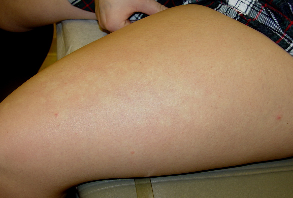
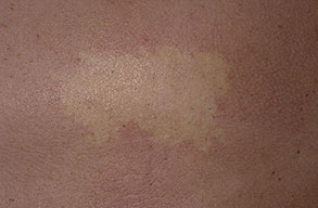
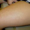
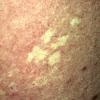
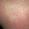
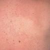
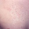
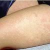
Nevus anemicus is a congenital vascular anomaly that presents clinically as a hypopigmented macule or patch, as shown below. The lesional pallor is due to a localized hypersensitivity to catecholamines with resultant vasoconstriction. Nevus anemicus is an uncommon disorder and was first described by Vorner in 1906.
Pathophysiology
Intralesional injection of bradykinin, acetylcholine, serotonin, nicotine, 5-hydroxytryptamine, and histamine fails to induce the anticipated vasodilatation or erythema in the affected area. However, erythema does follow an axillary sympathetic block or intradermal injection of the alpha-adrenergic blocking agent, pilocarpine. These findings suggest that nevus anemicus is best termed a pharmacologic nevus resulting from increased vascular sensitivity to catecholamines.1 This conclusion is further supported by autograft exchange transplantation studies that show donor site dominance. It also has been proposed that an abnormality in endothelial adhesion molecule induction (E selectin expression) may be involved, suggesting several pharmacologic anomalies are involved and further supporting the idea that nevus anemicus may best be termed a pharmacologic nevus.
The prevalence of nevus anemicus is not known, but it is not rare.
Lesions of nevus anemicus usually persist unchanged throughout life. They are asymptomatic.
No racial predilection has been noted in the literature for nevus anemicus.
Nevus anemicus appears more frequently in females.
Nevus anemicus may be present at birth or first appreciated in early childhood.Patients with nevus anemicus typically present with an asymptomatic pale macule or patch that has been present since birth and grows with the child. Frequently, the lesion of nevus anemicus is noted as an incidental finding on skin examination.
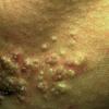
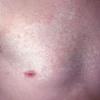
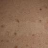
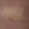
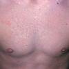
Clinically, nevus anemicus is a circumscribed, rounded, oval or linear pale macule or patch with irregular margins that may be surrounded by satellite macules. Lesions may be single or multiple and may be located on any part of the body, but most lesions commonly are found on the upper chest. Nevi anemicus occur more frequently in females and are usually asymptomatic. Nevus anemicus is noted at birth or in early childhood, although it may be easily overlooked.
Nevus anemicus usually persists unchanged throughout life. Lesions occur with increased frequency in patients with neurofibromatosis. Areas of nevus anemicus frequently are extensive and have been observed in close association with capillary malformations of port-wine stain type, a phenomenon attributed to somatic recombination. Nevus anemicus also has been described in patients with phakomatosis pigmentovascularis, a syndrome characterized by vascular and melanocytic nevi. Phakomatosis pigmentovascularis type IIa has been associated with primary choroidal melanoma.
Causes
Nevus anemicus is due to a congenital anomaly of the cutaneous vasculature resulting in hypersensitivity to catecholamines, leading to localized vasoconstriction.
Treatment
Medical Care
Treatment generally is not required for nevus anemicus. Patients troubled by the cosmetic appearance of the nevus anemicus lesion may benefit from the application of camouflage makeup.
Medication
No therapy is available for nevus anemicus.