|
synovial cyst = الكيسة المصلية |
 |
 |
 |
synovial cyst
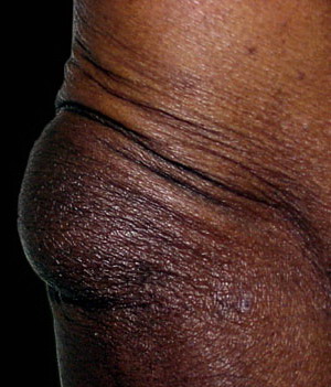
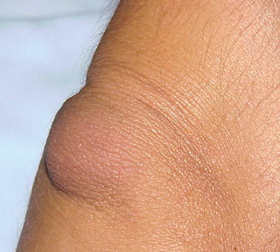
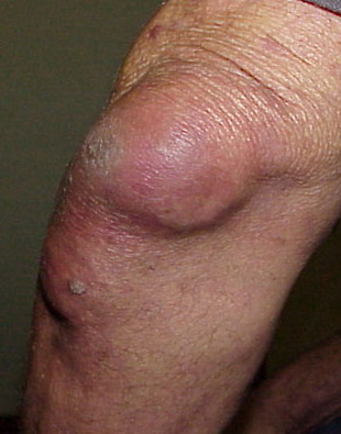
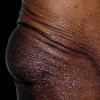
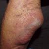
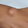
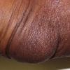
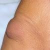
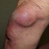
synovial cyst is a small, fluid-filled sac or pouch that can develop over a tendon or joint, creating a mass under the skin. Synovial cysts are found most commonly in the knee and hip. Cysts also can form in the shoulder, elbow, wrist (flexor tendon sheath in the fingers), top of the foot (dorsum), ankle, and hand. A synovial cyst may or may not be painful, depending on their size and location.
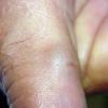
Ganglionic cysts are the most common cysts found on the on the hands and feet. A mucus (myxoid) cyst is a synovial cyst of the last joint of a digit (distal interphalangeal [DIP] joint). Myxoid cysts are thought to be caused by leakage of synovial fluid from the DIP joint, with or without underlying degenerative joint disease.
Synovial cysts may also develop in a degenerated spine, most frequently between the fourth and fifth lumbar vertebrae (L4-L5) where there is the most movement and potential for chronic trauma and instability. This type of cyst may limit spinal joint movement and compress adjacent nerves and vascular structures.
A Baker's cyst (popliteal cyst) forms in the back of the knee when fluid from inside the knee joint becomes trapped outside the joint. It can be caused by any condition that causes knee swelling (e.g., osteoarthritis, rheumatoid arthritis, meniscal tear).
Many times the cause of a synovial cyst is unknown (idiopathic), although there is some evidence that trauma may be a factor. The size of the sac or cyst can change with activity and may disappear for some time, only to recur. The mass is often soft, but with time it may become firmer to the touch.
Risk: Risk factors for developing synovial cysts are osteoarthritis, rheumatoid arthritis, acute or chronic trauma, joint instability, and overuse injuries from repetitive movement. Risk is increased for individuals who participate in repetitive activities such as keyboarding or playing a musical instrument (Cassidy).
Ganglion cysts of the hand and wrist are most common in women aged 20 to 30 (Cassidy).
Painful lumbar synovial cysts occur slightly more often in females than in males and are typically found in individuals in their seventh decade (Epstein, Khan).
Incidence and Prevalence: Ganglion cysts of the wrist and hand comprise 50% to 70% of all masses found in the hand; 80% of such cysts are located on the back of the hand (dorsal surface) (Cassidy).
Popliteal cysts are found in up to 20% of asymptomatic knees (Tschirch).
Incidence of lumbar synovial cysts is less than 0.5% of individuals with back pain in the general population (Khan).
|
History: Usually, the individual cannot pinpoint a significant incident that precipitated cyst formation; however, the individual will notice the cyst, whether painful or not, on the limbs, especially the ankle, wrist, fingers, or top of foot. Although pain may be present as the cyst forms, the pain usually goes away and the cyst becomes asymptomatic after a few months. The individual may report pain and swelling with activity, and the cyst may change in size with increased activity. Rarely, if a synovial cyst presses on a nerve, the individual may report feelings of tingling or numbness in the affected limb.
Physical exam: Palpation of the cyst reveals a soft to rigid mass near a joint or over a tendon. A ganglion cyst in the hand or wrist may reduce grip strength. With a myxoid cyst of the finger, the fingernail may appear deformed. With a popliteal cyst, the physician may palpate a thickening or soft tissue mass at the back of the affected knee. A synovial cyst that cannot be seen (occult) should be suspected for wrist pain without trauma. Although synovial cysts of the lumbar spine cannot be seen or palpated, they can result in muscle and sensory deficits and reflex changes.
Tests: Draining (aspirating) the cyst produces a thick, gelatinous liquid. X-rays may be ordered to rule out tumor or rheumatoid arthritis. Although not usually necessary, MRI is very useful in visualizing the cyst. In the case of occult synovial cysts of the lumbar spine, CT scan typically shows a soft tissue mass adjacent to a degenerative joint. A synovial cyst of the knee usually is not visible on plain x-rays, although x-rays can confirm the presence of osteoarthritis, which is associated with these cysts. An ultrasound or MRI is necessary to confirm the diagnosis of a popliteal cyst.
|
Conservative treatment for synovial cysts may include ice packs applied directly to the affected region together with oral medication for pain (e.g., acetaminophen, ibuprofen). Another form of conservative treatment for synovial cysts consists of aspirating the fluid from the sac with a large-gauge needle. The sac is then injected with a corticosteroid drug to shrink or dissolve it. With lumbar synovial cysts, steroid injections into the affected facet joint may be indicated. In longstanding, painful, or recurrent cases, techniques including fluoroscopically guided removal of the cyst, surgery to remove the cyst (minimally invasive excision) and its attachment to the tendon or joint capsule (stalk), or surgery to enlarge the space around the affected nerve root (laminectomy or foraminectomy) may be necessary (Sehati). The cyst also may spontaneously rupture, providing relief.
|
Symptoms of untreated symptomatic cysts worsen with increased activity. Aspiration followed by corticosteroid injection results in relief of symptoms in 35% of cases, but it is not uncommon for the cyst to return. Following minimally invasive surgical excision for a lumbar synovial cyst, 95% of cases obtain full resolution of symptoms (Sehati). Surgical removal provides complete relief of symptoms in most cases, but about 10% of cysts recur after surgery. Surgical removal of a myxoid cyst is more successful in the fingers than in the toes, with a cure rate of 95% for fingers but only 33% after toe surgery (Lawrence). In rare cases in which a cyst is cancerous, the prognosis will depend on the type of cancer and whether it has spread (metastasized).
|
Synovial cysts on weight-bearing surfaces of the foot may make standing or walking painful. Synovial cysts on the palm of the hand or at the wrist may interfere with grasping. Synovial cysts on the top of the foot or ankle may be irritated by shoe pressure.
If the synovial cyst involves the spine, neurological deficits such as numbness and muscle weakness can significantly complicate the condition and recovery. If the individual requires surgical excision, there may be complications associated with surgery (e.g., anesthesia, poor wound healing, infection). In case of a lumbar cyst, rarely further surgery may be needed to stabilize an unstable spine (lumbar fusion).
|
|
|
|
|