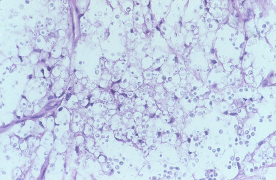
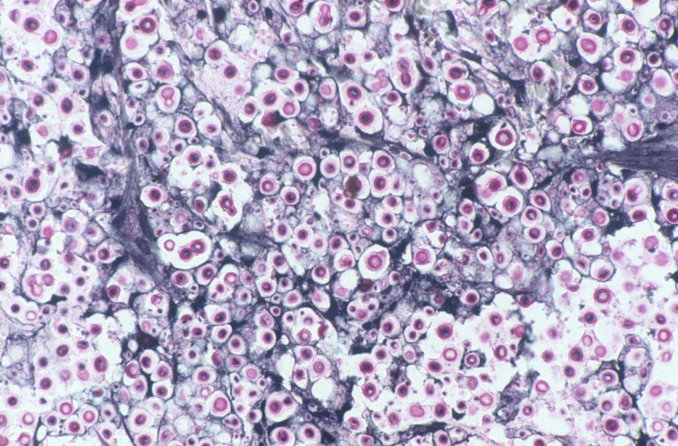
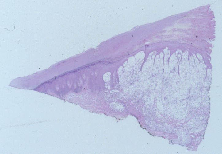
Histologic FindingsIn spinal fluid, urine, and tissue, pathogenic strains of C neoformans grow as round-to-oval yeast, surrounded by a polysaccharide capsule composed of mannose, xylose, and glucuronic acid. The yeast may be single or may have a single budding daughter cell. Cell size varies widely and ranges from 3.5-8 µm in diameter. Rarely, pseudohyphae develop. India ink, which outlines the organisms by negative contrast, helps to identify the yeast cells in fluids or macerated tissue samples. In fixed tissue, the capsule of C neoformans may also be stained with mucicarmine, which preferentially stains mucopolysaccharides. Tissue sections can be stained with the Fontana-Masson stain to detect melanin precursors in the yeast cell wall. The presence of melanin or melanin precursors is useful in differentiating C neoformans from other yeasts. |