|
Scleromyxedema = الوذمة المخاطية الصلبة |
 |
|
Scleromyxedema
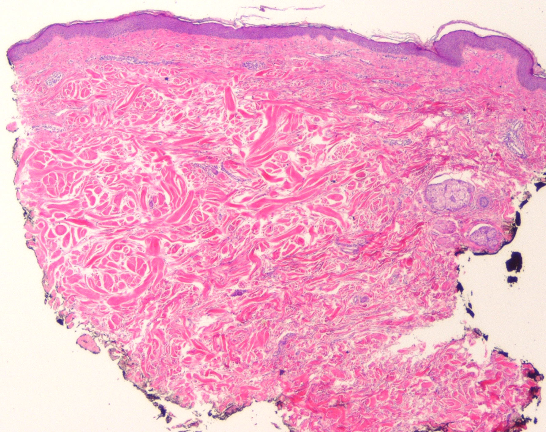
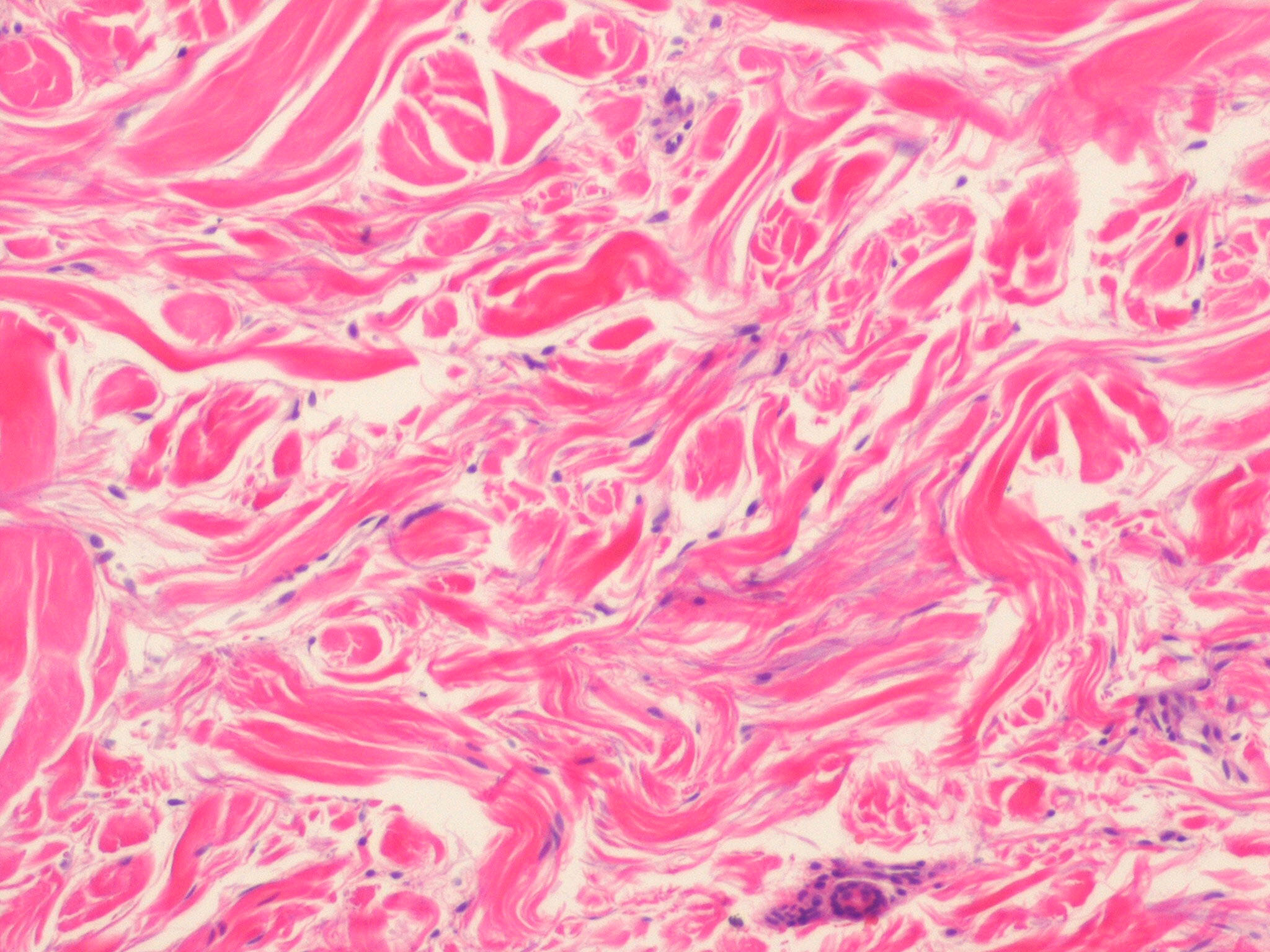
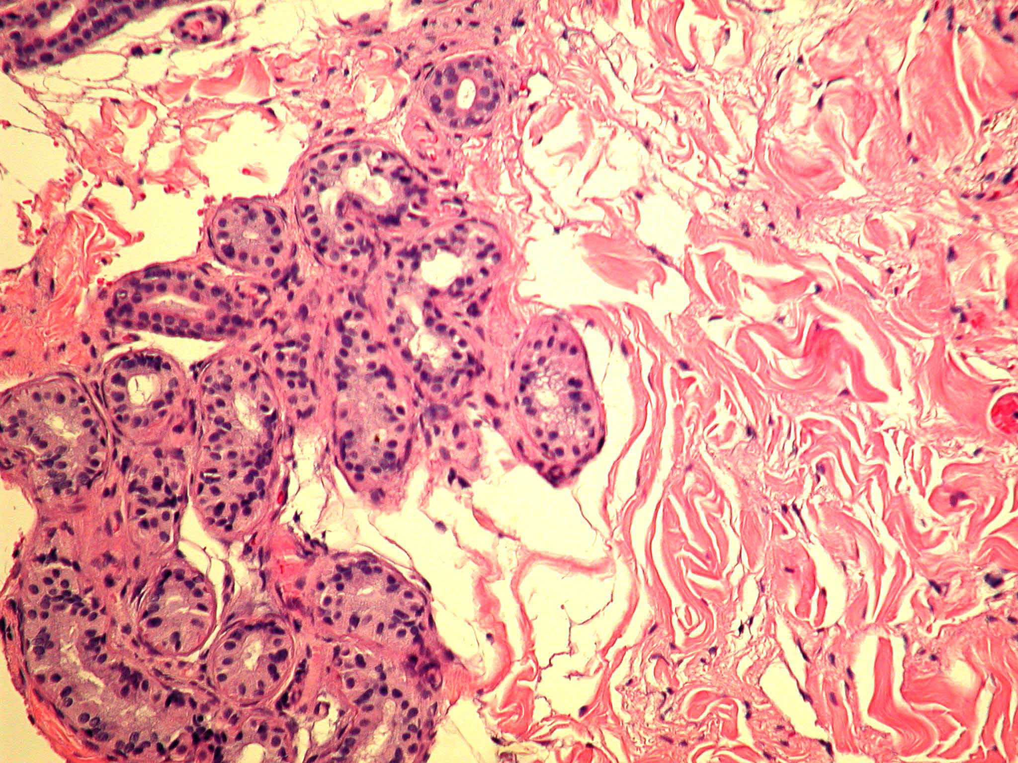
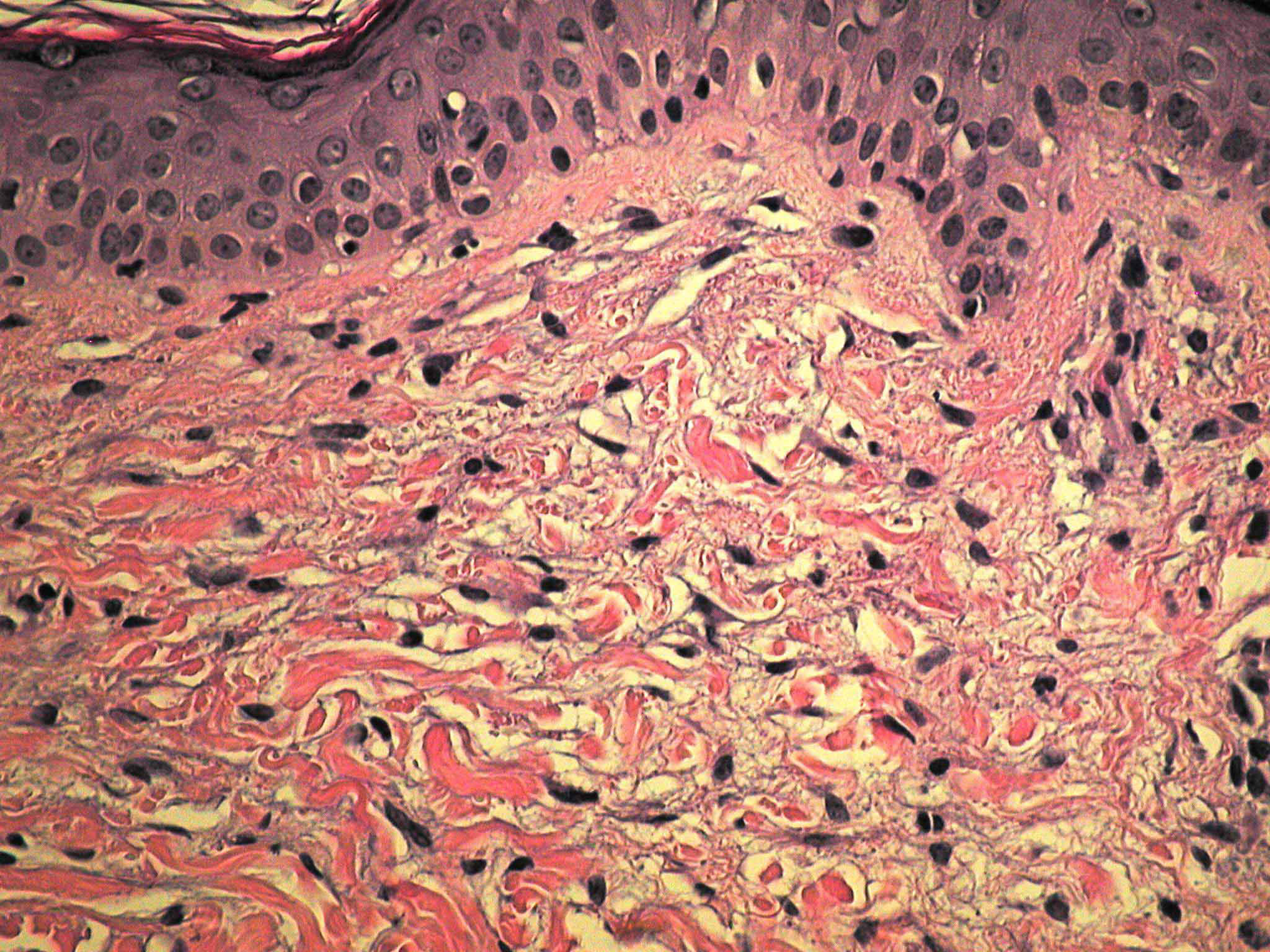
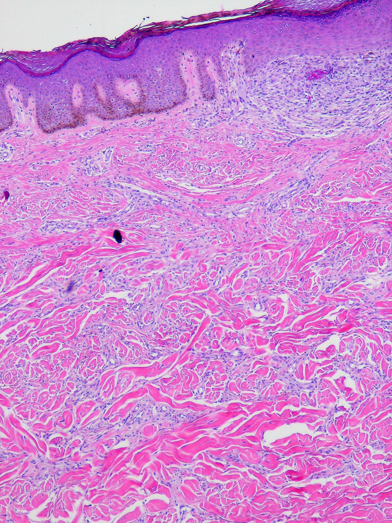
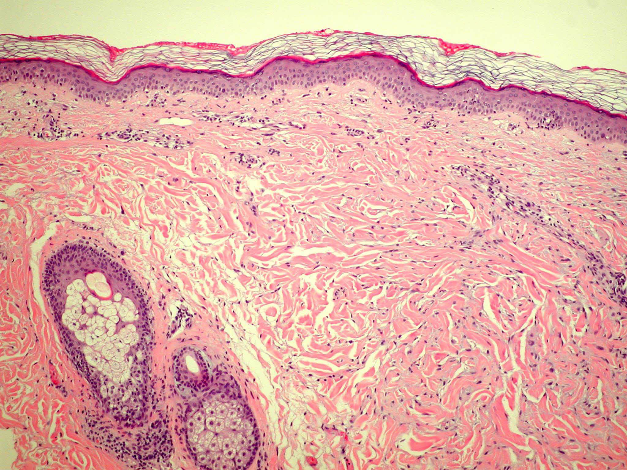
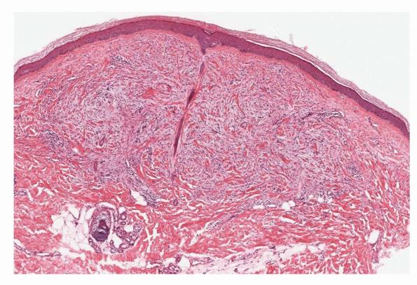
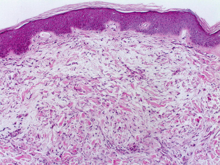
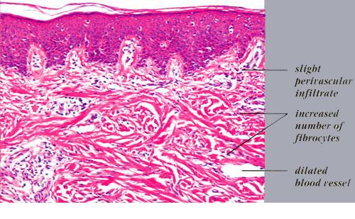
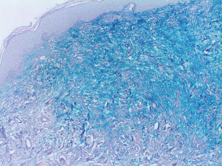
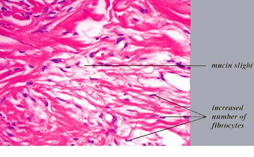
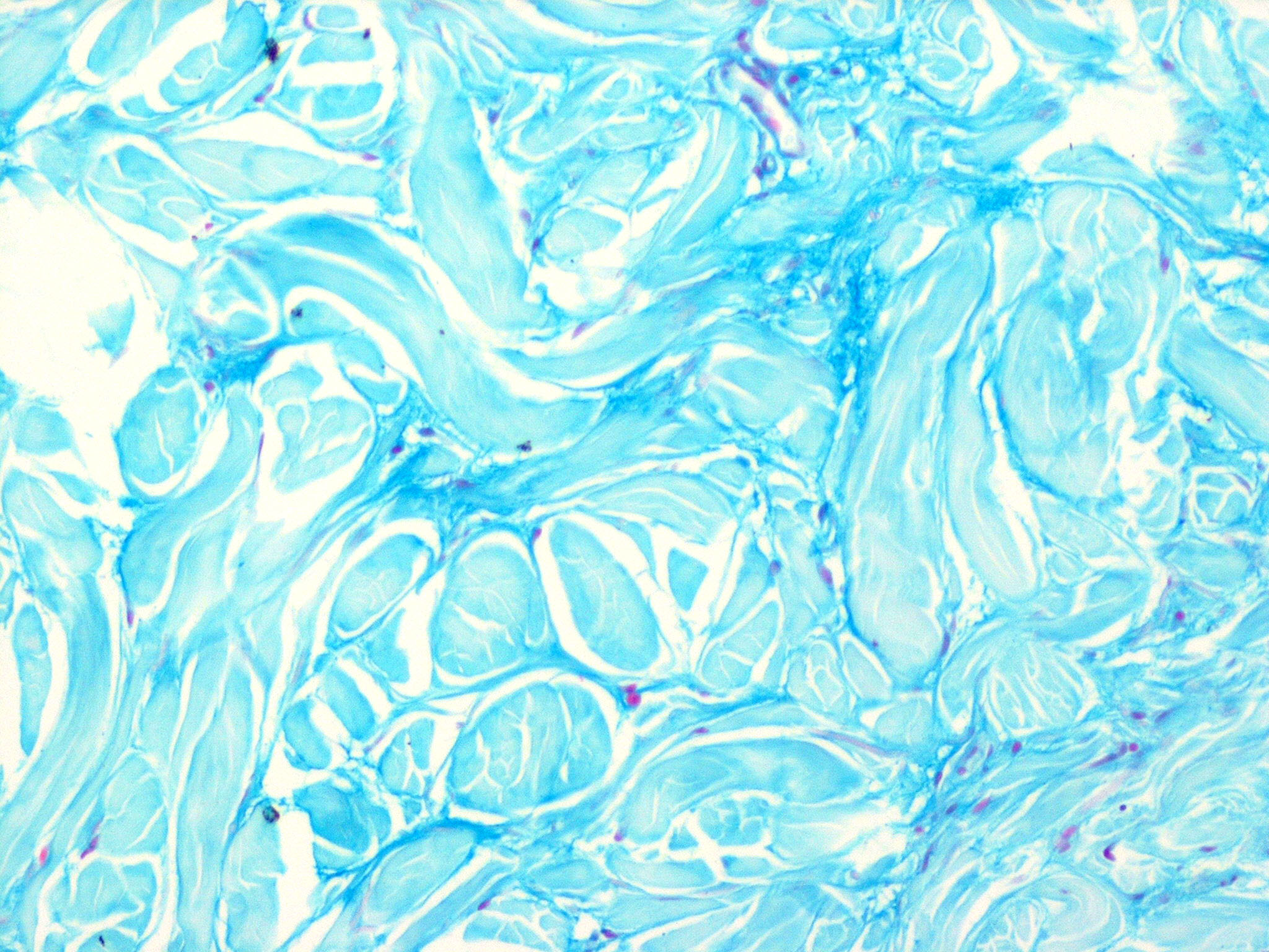
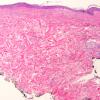
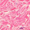
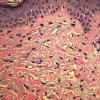
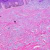
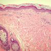
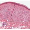
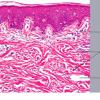
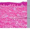
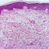
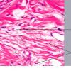
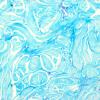
The histologic picture found in the papules in scleromyxedema resembles that observed in lichen myxedematosus. In the diffusely thickened skin, there is extensive proliferation of fibroblasts throughout the dermis associated with irregularly arranged bundles of collagen. In many areas, the collagen bundles are split into individual fibers by mucin. As a rule, the amount of mucin is greater in the upper half than in the lower half of the dermis.
|
|
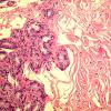
Scleromyxedema may be difficult to distinguish from nephrogenic fibrosing dermopathy without clinicopathologic correlation. Both conditions are characterized by a dermal infiltrate of spindle cells with increased mucin and collagen. In both conditions, the spindle cells label for either factor Xilla or CD34 and procollagen1 without demonstrating a pattern that could be utilized to discriminate between these diseases . However, in scleromyxedema, the infiltrate is limited to the dermis whereas in nephrogenic fibrosing dermopathy the infiltrate usually also extends down the subcutaneous septa .
|
|
Autopsy examination of patients with lichen myxedematosus and scleromyxedema usually has not shown mucinous deposits in any internal organs . However, in one case, mucin was detected in renal papillae, and in others, it was found in the adventitia and media of blood vessels in several organs .
|
|
Pathogenesis. Electron microscopic examination of scleromyxedema reveals an increase in the number of fibroblasts. They show considerable activity, as indicated by the presence of a markedly dilated rough endoplasmic reticulum and long cytoplasmic processes. These fibroblasts produce both collagen and ground substance. The presence of many collagen fibrils with reduced diameter, similar to those in scleroderma, indicates that it is young collagen. In many areas, there are small bundles of young collagen fibrils, with each fibril richly coated with ground substance .
|
|
The presence of a monoclonal component (M component) or paraprotein in the sera of patients with lichen myxedematosus and scleromyxedema has been noted in nearly all cases that have been adequately tested. Its absence has been noted in a few cases ; in another case, it was absent at first but present later on .
|
|
In nearly all instances, the paraprotein is an IgG, although other immunoglobulin classes occur on rare occasions . The IgG paraprotein is a very basic protein because of an increased lysine content in its light chains . Thus, in most instances, it migrates more slowly than gamma globulin on serum electrophoresis and may even migrate toward the cathode rather than toward the anode . In instances in which it migrates with the same speed as gamma globulin, immunoelectrophoresis is required for its recognition . The IgG of lichen myxedematosus differs from the IgG of multiple myeloma not only by usually showing slower electrophoretic migration but also by the fact that its IgG molecules nearly always possess light chains of the lambda type, although light chains of the kappa type have occasionally been found . In contrast, in multiple myeloma with elevated values for IgG, only about one third of the reported cases with IgG molecules have lambda light chains, and two thirds have kappa light chains . In addition to showing a monoclonallgG, many cases of lichen myxedematosus show hyperplasia of plasma cells in the bone marrow . These plasma cells have been shown to synthesize the monoclonallgG . In some cases, the plasma cells in the bone marrow have been regarded as atypical in appearance . However, only one case fulfilling the criteria for coexisting multiple myeloma has been documented .
|
|
The role of the monoclonal IgG in lichen myxedematosus is not clear. Its presence in the dermal mucin has been demonstrated by direct immunofluorescence . Furthermore, the serum containing the paraprotein stimulates the production of hyaluronic acid and prostaglandin E by fibroblasts in vitro . However, although the serum from patients with lichen myxedematosus stimulates fibroblast proliferation in vitro, the purified IgG paraprotein itself has no direct effect on fibroblast proliferation .
|
|