|
Papular mucinosis = الداء المخاطيني الحطاطي |
 |
|
Mucinoses
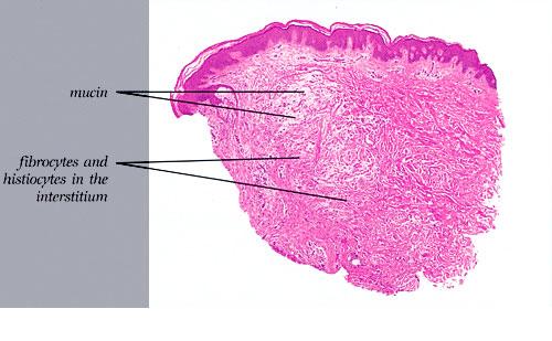
There are six types of cutaneous mucinosis: (a) generalized myxedema, (b) pretibial myxedema, (c) lichen myxedematosus or papular mucinosis, (d) reticular erythematous mucinosis or plaquelike mucinosis, (e) selfhealing juvenile cutaneous mucinosis, and (f) scleredema.
|
|
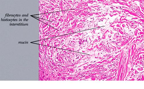
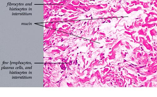
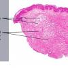
Regular demonstration of the presence of mucin in the dermis is possible only in pretibial myxedema, self-healing juvenile cutaneous mucinosis, and lichen myxedematosus. In reticular erythematous mucinosis, it is possible in most cases. In generalized myxedema, the amount of mucin usually is too small to be demonstrable, and in scleredema, mucin may be present only in the early stage.
|
|
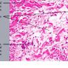
The mucin found in these six diseases represents an increase in the mucin that is normally present in the ground substance of the dermis. It consists of proteins bound to hyaluronic acid (hyaluronan), which is an acid mucopolysaccharide or glycosaminoglycan. As a result of the great water-binding capacity of hyaluronic acid, dermal mucin contains a considerable amount of water. This water is largely removed during the process of dehydration of the specimen; consequently, in routine sections, the mucin, because of its marked shrinkage, appears largely as threads and granules.
|
|
The mucin present in the six types of mucinosis stains a light blue in sections stained with H&E. It also stains with colloidal iron. It is Alcian blue-positive at pH 2.5 but negative at pH 0.5 and shows metachromasia with toluidine blue at pH 7.0 and 4.0 but no metachromasia below pH 2.0 . It is PAS negative (indicating the absence of neutral mucopolysaccharides) and aldehyde fuchsin negative (indicating the absence of sulfated acid mucopolysaccharides). The mucin is completely removed on incubation of histologic sections with testicular hyaluronidase for 1 hour at 37"C .
|
|