| Lichen nitidus = الحزاز الشوكي |
|
|
Lichen Nitidus
Lichen nitidus (Latin nitidus, “shiny” or “glistening”) is an uncommon, usually asymptomatic cutaneous eruption first described by Felix Pinkus in 1901 and further characterized by him in 1907. The dermatosis consists of small, glistening, flesh-colored to pink or reddish-brown papules that may be limited to the penis, genitalia, abdomen, and extremities or, less frequently, may occur as a generalized condition. The histopathologic findings are characteristic. Although the condition is often chronic, the prognosis is good, and no clearly associated systemic illnesses have been documented.
EPIDEMIOLOGY Because the disease is uncommon, accurate epidemiologic characteristics of lichen nitidus have not yet been defined. Lichen nitidus is a dermatosis that occurs infrequently and has been reported to affect blacks more than Caucasians, although no strong predisposition for any race is clearly evident. Perhaps the relative prominence of pale lesions on dark skin accounts for the reported increased incidence in blacks. A predilection for children, young adults, and males has also been reported, but, again, these data are not well established. The incidence is estimated to be approximately 3.4 cases/10,000 population, based on a 25-year survey of skin diseases in African Americans. Compared to the more common lichen planus, the crude ratio of lichen nitidus to lichen planus is 1.7:100, based on pathologic diagnosis of cases evaluated over several decades at the Mayo Clinic. ▪ ETIOLOGY AND PATHOGENESIS Once considered a tuberculoid reaction, lichen nitidus is currently regarded as a disorder of unknown etiology. The relationship between lichen nitidus and lichen planus has been debated for many
LICHEN NITIDUS AT A GLANCE
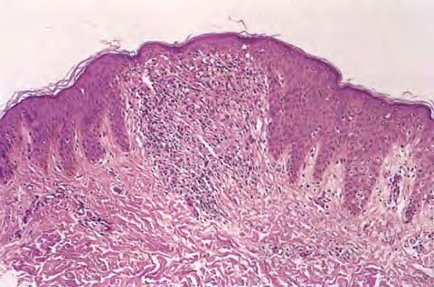
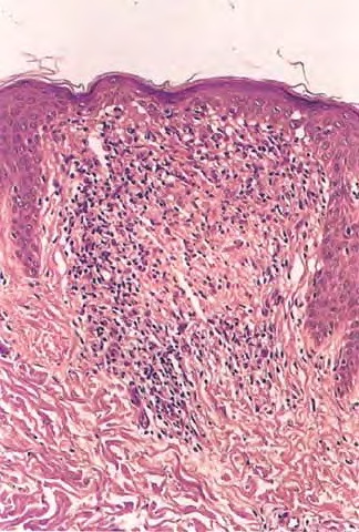
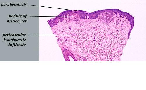
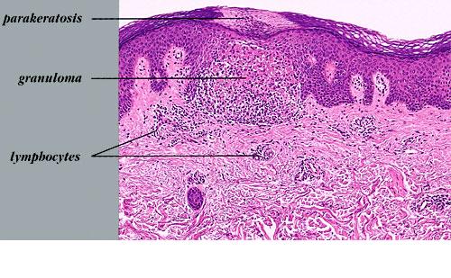
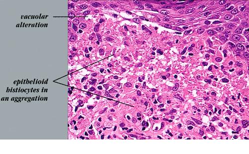
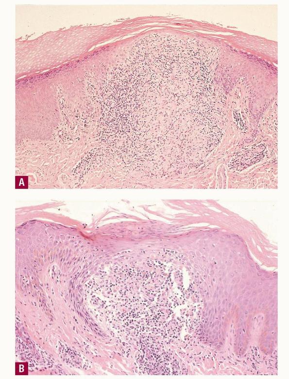
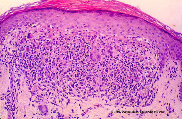
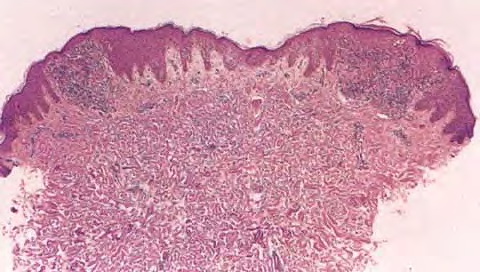
A rare familial presentation of lichen nitidus has been reported, although no genetic factors of the disease have been identified.9 CLINICAL FINDINGS Lichen nitidus is composed of multiple, discrete, smooth, flat, round papules. Individual papules are 1 to 2 mm in size, flesh-colored to slightly pink or, in blacks, hypopigmented, with a glistening appearance . Sometimes, minimal scale is present or can be elicited by rubbing the surface of the papules. Occasionally, the papules are grouped and the isomorphic or Koebner phenomenon is observed. Lesions may occur anywhere over the skin surface; however, the most frequent sites of predilection are the flexural surfaces of the arms and the wrists, lower abdomen, breasts, the glans and shaft of the penis, and other areas of the genital region. Rare sites of involvement include mucous membranes, nails, palms, and soles. Rare clinical variants include vesicular, hemorrhagic, spinous follicular, linear, generalized, and actinic types. Palmo-plantar involvement may manifest several morphologic forms. Bilateral hyperkeratosis of palms and soles with erythema, fissuring, and a texture resembling fine sandpaper has been observed. Occasionally, minute keratotic spicules on the palmar surfaces or multiple pinpoint papules that extend to the dorsa of the extremities occur . Nail abnormalities usually manifest as longitudinal, beaded ridging, and terminal splitting with or without irregular pitting. Lesions of lichen planus may infrequently be present simultaneously. Lichen nitidus is usually asymptomatic; however, pruritus is occasionally present and sometimes intense. There are no constitutional symptoms or systemic abnormalities associated with the disease. Lichen nitidus has been reported after hepatitis B vaccination.18 A dense mass of infiltrating lymphohistiocytic cells is situated immediately below the epidermis and results in widening of the papillary dermis with elongation and the appearance of embracement by neighboring rete ridges . Occasionally, two or three papillary spaces merge together as part of the inflammatory
The majority of the cells in the infiltrate are T lymphocytes intermixed
Direct immunofluorescence examination of lichen nitidus is usually negative for deposition of immunoglobulins at the dermal-epidermal junction, in contrast to the vast majority (95 percent) of cases of lichen planus . Cytoids are also not usually observed in lichen nitidus. The results of ultrastructural studies coincide with light microscopic findings and also show activated lymphocyte morphology with convoluted nuclei, resembling Sézary cells. PROGNOSIS AND CLINICAL COURSE Lichen nitidus is typically a focal, asymptomatic, chronic inflammatory reaction that eventually resolves spontaneously after months to 1 year in two-thirds of patients or, less frequently, over a few years. Rarely, the eruption may persist indefinitely. New lesions may continue to develop as older lesions resolve. Lesions heal without scar formation or pigmentary abnormalities. Differential Diagnosis of Lichen Nitidus Most Likely
Consider
TREATMENT Because the disease is asymptomatic and self-limiting, no intervention is required in most cases. Treatment of lichen nitidus is warranted when it is associated with protracted pruritus or when the appearance interferes with the patient's daily activities and outlook. Topical glucocorticoids may yield favorable results. A short course of oral glucocorticoids may also be helpful and hasten resolution of more extensive, generalized, or symptomatic disease.Psoralen and ultraviolet A light, ultraviolet A and B phototherapy, astemizole, acitretin or etretinate, low-dose cyclosporine,and oral itraconazole have also been used successfully when indicated for more problematic disease. Narrowband ultraviolet B could be a safe and effective treatment in generalized lichen nitidus. Tacrolimus ointment was also reported to be effective after 1 month of therapy.History of exposure to tuberculosis in the setting of lichen nitidus should be investigated and when appropriate treated with antituberculous medications. Complete clearance of lichen nitidus was seen in one case.
|
|||||||||||||||||||||||||||||||||||||||||||||||||||||||||||||||||||||||||||||||||||||||||||||||