|
Grover's disease_transient acantholytic dermatosis = داء غروفر =جلاد انحلال الاشواك العابر |
 |
 |
 |
|
Transient Acantholytic Dermatosis Grovers Disease
First described in 1970, transient acantholytic dermatosis is characterized by pruritic, discrete papules and papulovesicles on the chest, back, and thighs. In rare instances, vesicles and even bullae are seen. Most patients are middle-aged or elderly men. Although the disorder is transient in the majority of patients, lasting from 2 weeks to 3 months, it can persist for several years. The condition has been reported coexisting with other dermatosis, such as asteatotic eczema, allergic contact dermatitis, atopic eczema, psoriasis, and pemphigus foliaceus. There have been reports of patients with transient acantholytic dermatosis and malignancy, most commonly lymphoproliferative and genitourinary neoplasms .
|
|
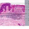
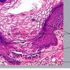
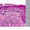
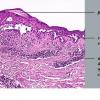
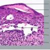
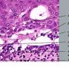
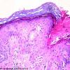
Histopathology.
Focal acantholysis and dyskeratosis (focal acantholytic dyskeratosis) are present. Because these foci are small, they are sometimes found only when serial sections are obtained. The acantholysis may occur in four histologic patterns, resembling Darier's disease , Hailey-Hailey disease, pemphigus vulgaris, or spongiotic dermatitis. Two or more of these patterns may be found in the same specimen. There is usually a superficial dermal infiltrate of lymphocytes and sometimes eosinophils.
|
|
IF Testing. In general, IF results are often said to be negative. However, nonspecific granular basement membrane zone staining of C3 alone or in combination with IgM may be found.
|
|
Pathogenesis. Despite the histologic similarity to Darier's disease, Grover's disease does not share an abnormality in the ATP2A2 gene , which encodes a keratinocyte Ca2+ pump. Mutation of ATP2A2 is responsible for Darier's disease. Although the pathogenesis of Grover's disease remains unknown, there appears to be a relationship to excessive sweating, fever, and bed confinement. Some authors have hypothesized that heat or sweat urea that leaks from the intraepidermal portion of the sweat duct into the surrounding epidermis causes acantholysis. Others dispute this theory and have found the sweat duct to be intact. Finally, IL-4 may be responsible for acantholysis, either by induction of plasminogen activator or by stimulation of antibody production . The expression of syndecan-1, a proteoglycan important for keratinocytes intercellular adhesion, is markedly
|
|
Ultrastructural Study. In the pemphigus-like zones, there is intradesmosomal separation, fewer desmosomes, and perinuclear aggregation of tonofilament bundles. In the Darier type, features similar to those of Darier's disease are present.
|
|
Differential Diagnosis. The features that help to differentiate transient acantholytic dermatosis from the four diseases that it resembles are the focal nature of the histologic changes and the mixture of patterns. The presence of eosinophils in the superficial dermal infiltrate of Grover's disease serves as a distinguishing feature from Darier's disease, in which they are usually absent. It may be important to have clinical information for a definitive diagnosis. IF studies are rarely necessary to exclude pemphigus.
|
|

