|
Epidermal cyst = الكيسة البشروية |
 |
 |
 |
|
Histopathology
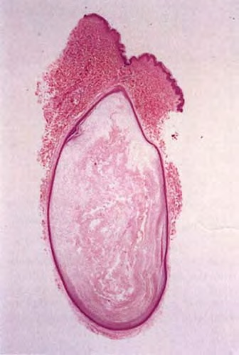
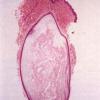
Epidermal cysts have a wall composed of true epidermis, as seen on the skin surface and in the infundibulum of hair follicles, the infundibulum being the uppermost part of the hair follicle that extends down to the entry of the sebaceous duct. In young epidermal cysts, several layers of squamous
and granular cells can usually be recognized . In older epidermal cysts, the wall often is markedly atrophic, either in some areas or in the entire cyst, and may consist of only one or two rows of greatly flattened cells. The cyst is filled with horny material arranged in laminated layers. In sections stained with hematoxylin-eosin, melanocytes and melanin pigmentation of keratinocytes can be seen only rarely in epidermal cysts of whites but frequently in epidermal cysts of blacks. Silver stains reveal that most of the melanin is located in the basal layer of the cyst lining, but some melanin is seen also in the contents of the cyst .
|
|
When an epidermal cyst ruptures and the contents of the cyst are released into the dermis, a considerable foreign-body reaction with numerous multinucleated giant cells results, forming a keratin granuloma . The foreign-body reaction usually causes disintegration of the cyst wall. However, it may lead to a pseudocarcinomatous proliferation in remnants of the cyst wall, simulating a squamous cell
care i nom a . Mel a n 0 cyt e s, mel ani n, and melan0phag e s I have been reported in the walls of
epidermal cysts in some Indians and a Japanese patient
|
|
having reached 1 to 5 em in diameter. They occur most commonly on the face, scalp, neck, and trunk . Although most epidermal cysts arise spontaneously in hair-bearing areas, occasionally they occur on the palms or soles or form as the result of trauma (137). Usually a patient has only one or a few epidermal cysts, rarely many. In Gardner's syndrome, however, numerous epidermal cysts occur, especially on the scalp and face .
|
|
Epidermal cysts are slowly growing, elevated, round, firm, intradermal or subcutaneous tumors
|
|
Development of a basal cell epithelioma , a lesion of Bowen's disease , or a squamous cell carcinoma in epidermal cysts is a rare event. In cases of squamous cell carcinoma, the tumor is apt to be of low malignancy and does not metastasize. It is likely that some cases that were regarded in the past as malignant degeneration of epidermal cysts now are interpreted either as pseudocarcinomatous hyperplasia in a ruptured epidermal cyst or as proliferating trichilemmal tumor .
|
|
Pathogenesis. It is widely assumed that most spontaneously arising epidermal cysts are related to the follicular infundibulum. The occurrence of hybrid cysts with partially epidermal and partially trichilemmal lining favors this assumption . Epidermal cysts in nonfollicular regions, such as the palms or soles, probably form as a result of the traumatic implantation of epidermis into the dermis or subcutis .
|
|
have been reported in the walls of epidermal cysts in some Indians and a Japanese patient .
|
|
Development of a basal cell epithelioma , a lesion of Bowen's disease , or a squamous cell carcinoma in epidermal cysts is a rare event. In cases of squamous cell carcinoma, the tumor is apt to be of low malignancy and does not metastasize. It is likely that some cases that were regarded in the past as malignant degeneration of epidermal cysts now are interpreted either as pseudocarcinomatous hyperplasia in a ruptured epidermal cyst or as proliferating trichilemmal tumor .
|
|
Pathogenesis
It is widely assumed that most spontaneously arising epidermal cysts are related to the follicular infundibulum. The occurrence of hybrid cysts with partially epidermal and partially trichilemmal lining favors this assumption . Epidermal cysts in nonfollicular regions, such as the palms or soles, probably form as a result of the traumatic implantation of epidermis into the dermis or subcutis .
|
As seen by electron microscopy, the keratinization in epidermal cysts is identical to that in the surface epidermis and in the pilosebaceous infundibulum because the keratin located within the keratinized cells consists of relatively electron-lucent tonofilaments embedded in an electron-dense interfilamentous substance derived from keratohyaline granules. The keratinized cells of the cyst content have a markedly flattened, elongated appearance and are surrounded by a thick marginal band rather than by a plasma membrane. Desmosomes are no longer present
|