| Bullous erythema multiforme =الحمامى عديدة الأشكال الفقاعية |
|
|
Erythema Multiforme
Erythema multiforme (EM) is an acute self-limited, usually mild, and often relapsing mucocutaneous syndrome. The disease is usually related to an acute infection, most often a recurrent herpes simplex virus (HSV) infection. EM is defined only by its clinical characteristics: target-shaped plaques predominant on the face and extremities.
The absence of specific pathology, specific cause, and biologic markers contribute to a confusing nosology. The definition of EM in this chapter is based on the classification proposed by Bastuji-Garin et al. The principle of this classification is to consider Stevens-Johnson syndrome (SJS) as a minor form of toxic epidermal necrolysis (TEN) and to separate these two diseases from EM . The validity of this classification has been challenged by some studies, especially for cases in children and cases related to Mycoplasma pneumoniae. It has been confirmed by several others studies, however, especially the prospective international Severe Cutaneous Adverse Reactions study.2 That study demonstrated that, compared with SJS patients, EM patients were younger, more often male, had a 10-fold higher rate of recurrence, and less frequently had a body temperature higher than 38.5°C (101.3°F) and involvement of two or more mucous membranes. The original name proposed by von Hebra was erythema exudativum multiforme. The term erythema multiforme has now been universally accepted (Table 38-1). EPIDEMIOLOGY EM is considered relatively common, but its incidence is unknown. Evaluations have been limited to cases severe enough to require hospitalization. Such cases are definitely rare, with figures in the range of 1 to 6 per million per year. Minor forms of EM are certainly more frequent, but the diagnosis of EM is nonetheless too often made.
ERYTHEMA MULTIFORME AT A GLANCE
EM occurs in patients of all ages, but mostly in adolescents and young adults. There is a slight male preponderance (male-female sex ratio of approximately 3:2). EM is recurrent in at least 30 percent of patients. There is no established underlying disease. Infection with human immunodeficiency
Predisposing genes have been reported, with 66 percent of EM patients having HLA-DQB1*0301 allele, compared with 31 percent of controls.3 The association was even stronger in patients with herpes-associated EM. Because of the weakness of this association, familial cases remain very rare. Most cases of EM are related to infections. Herpes virus is definitely the most common cause, principally in recurrent cases. Proof of causality of herpes is firmly established from clinical experience, epidemiology, detection of HSV DNA in the lesions of EM, and prevention of EM by suppression of HSV recurrences.Clinically, a link with herpes can be established in about one-half of cases. In addition 10 percent to 40 percent of cases without clinical suspicion of herpes were also demonstrated to be herpes related, because HSV DNA was detected in the EM lesions by polymerase chain reaction (PCR) testing. EM eruptions begin on average 7 days after a recurrence of herpes. The delay can be substantially shorter. Not all symptomatic herpes recurrences are followed by EM, and asymptomatic ones can induce EM. Therefore, this causality link can be overlooked by both patients and physicians. HSV-1 is usually the cause, but HSV-2 can also induce EM. The proportion probably reflects the prevalence of infection by HSV subtypes in the population. M. pneumoniae is the second major cause of EM and may even be the major cause in pediatric cases. In cases related to M. pneumoniae the clinical presentation is often less typical and more severe than in cases associated with HSV. The relationship to M. pneumoniae is often difficult to establish. Clinical and radiologic signs of atypical pneumonia can be mild, and M. pneumoniae is usually not directly detected. PCR testing of throat swabs is the most sensitive technique. Serologic results are considered diagnostic in the presence of immunoglobulin M antibodies or a more than twofold increase in immunoglobulin G antibodies to M. pneumoniae. M. pneumoniae-related EM can recur. Many other infections have been reported to be causes of EM in individual cases or small series, but the evidence for causality of these other agents is only circumstantial. Published reports have implicated infection with orf virus, varicella-zoster virus, parvovirus B19, and hepatitis B and C viruses, as well as infectious mononucleosis and a variety of other bacterial or viral infections. Immunization has been also implicated as a cause in children. Drugs are a rare cause of EM with mucous membrane lesions. It may be argued whether these eruptions are truly EM or mere imitators, for example, annular urticaria or maculopapular eruption with some lesions resembling targets. Idiopathic cases are those in which neither HSV infection nor any other cause can be identified. Such cases are fairly common under routine circumstances. But HSV has been found in situ by PCR in up to 40 percent of “idiopathic” recurrent cases.10 Some such cases respond to prophylactic antiviral treatment and are thus likely to have been triggered by asymptomatic HSV infection; others are resistant. EM-like dermatitis may result from contact sensitization. These rashes should be viewed as imitators of EM, despite their clinical and histopathologic similarities. PATHOGENESIS Mechanisms have been investigated in depth in herpes-associated EM. It is not known whether similar pathophysiologic mechanisms apply to EM due to other causes. Complete infective HSV has never been isolated from lesions of herpes-associated EM. The presence of HSV DNA in EM lesions has been reported in numerous studies using the PCR assay. These studies demonstrated that keratinocytes did not contain complete viral DNA but only fragments, always including the viral polymerase (Pol) gene. HSV Pol DNA is located in basal keratinocytes and in lower spinous cell layers. The viral Pol protein is synthesized in epidermal cells. HSV-specific T cells, including cytotoxic cells, are recruited, and the virus-specific response is followed by a non-specific inflammatory amplification by autoreactive T cells. These cells and the cytokines they produce induce the delayed hypersensitivity-like appearance of the pathology of EM lesions. HSV is present in the blood for a few days around an overt recurrence of herpes. If keratinocytes were infected in that way, one would expect disseminated herpes rather than EM. It has been shown that HSV DNA is transported to the epidermis by cells that engulf the virus and fragment the DNA. These cells are monocytes, macrophages, and especially CD34+ Langerhans cell progenitors harboring the skin homing receptor cutaneous lymphocyte-associated antigen. Upregulation of adhesion molecules greatly increases binding of HSV-containing mononuclear cells to endothelial cells. HLA-class I and adhesion molecule upregulation in endothelial cells may account for the dermal inflammatory response. When reaching the epidermis the cells transmit the viral Pol gene to keratinocytes. The genes may persist for a few months, but the synthesis and expression of the Pol protein will last for only a few days. This may explain the transient character of clinical lesions. Incomplete fragmentation of viral DNA, increased number of circulating CD34+ cells, and/or increased immune response to Pol protein may explain why only a small proportion of persons with recurrent herpes develop EM. CLINICAL FINDINGS The first step is to suspect EM, based on clinical features. A skin biopsy and laboratory investigations are useful mainly if the diagnosis is not definite clinically. The second step is to determine whether hospitalization is needed when EM major (EMM) occurs with oral lesions that impair feeding, when a possible diagnosis of SJS is suspected, or when severe constitutional symptoms are present. The third step is to establish the cause of EM by identifying a history of recurrent herpes, performing chest radiography, or documenting M. pneumoniae infection . History Prodromal symptoms are absent in most cases. If present, they are usually mild, suggesting an upper respiratory infection (e.g., cough, rhinitis, low-grade fever). In EMM, fever higher than 38.5°C (101.3°F) is present in one-third of cases. A history of previous attack(s) is found in at least one-third of patients and thus helps with the diagnosis. The events of the preceding 3 weeks should be reviewed for clinical evidence of any precipitating agent, with a special focus on recurrent herpes. Cutaneous Lesions The skin rash arises abruptly. In most patients, all lesions appear within 3 days, but in some, several crops follow each other during one episode of EM. Often there are a limited number of lesions, but up to hundreds may form. Most occur in a symmetric, acral distribution on the extensor surfaces of the extremities (hands and feet, elbows, and knees), face, and neck and appear less frequently on thighs, buttocks, and trunk. Lesions often first appear acrally and then spread in a centripetal manner. Mechanical factors (Koebner phenomenon) and actinic factors (predilection of sun-exposed sites) appear to influence the distribution of lesions. Although patients occasionally report burning and itching, the eruption is usually asymptomatic. The diversity in clinical pattern implied by the name multiforme is mainly due to the findings in each single lesion; most lesions are usually rather similar in a given patient at a given time. The typical lesion is a highly regular, circular, wheal-like erythematous papule or plaque that persists for 1 week or longer . It measures from a few millimeters to approximately 3 cm and may expand slightly over 24 to 48 hours. Although the periphery remains erythematous and edematous, the center becomes violaceous and dark; inflammatory activity may regress or relapse in the center, which gives rise to concentric rings of color . Often, the center turns purpuric and/or necrotic or transforms into a tense vesicle or bulla. The result is the classic target or iris lesion. According to the proposed classification, typical target lesions consist of at least three concentric components: (1) a dusky central disk, or blister; (2) more peripherally, an infiltrated pale ring; and (3) an erythematous halo. Not all lesions of EM are typical; some display two rings only (“raised atypical targets”). However, all are papular, in contrast with macules, which are the typical lesions in SJS-TEN. In some patients with EM, most lesions are livid vesicles overlying a just slightly darker central portion, encircled by an erythematous margin . Larger lesions may have a central bulla and a marginal ring of vesicles (herpes iris of Bateman) . Unusual presentations include cases in which recurrent EM in the same patient produces typical target lesions in one instance but plaques in a subsequent event. Mucous membranes can be severely involved in some episodes and spared in others (see Mucous Membrane Lesions). In most cases, EM affects well under 10 percent of the body surface area. In 88 hospital cases of EMM prospectively included in the Severe Cutaneous Adverse Reactions study, the median involvement was 1 percent of the body surface area.2 Very rare instances of extensive skin lesions with “giant” targets and prominent involvement of several mucous sites may be difficult to distinguish from SJS. The duration of an individual lesion is shorter than 2 weeks, but residual pigmentation may remain for months. There is no scarring. Mucous Membrane Lesions Mucosal lesions are present in up to 70 percent of patients, most often limited to the oral cavity. Predilection sites for mucosal lesions are the lips, on both cutaneous and mucosal sides; non-attached gingivae; and the ventral side of the tongue. The hard palate is usually spared, as are the attached gingivae. On the cutaneous part of the lips, identifiable target lesions may be discernible (see Fig. 38-3). On the mucosa proper there are erosions with fibrinous deposits, and occasionally intact vesicles and bullae can be seen . The process may rarely extend to the throat, larynx, and even the trachea and bronchi. Eye involvement begins with pain and bilateral conjunctivitis in which vesicles and erosions can occur . The nasal, urethral, and anal mucosae also may be inflamed and eroded. Ectodermosis pluriorificialis is a rare occurrence characterized by severe involvement of two or three mucosal sites in the absence of skin lesions. Its often relapsing nature suggests that it is HSV related. Moreover, typical target lesions may arise on the skin with new attacks. Relationship to Recurrent Herpes In more than 70 percent of patients with recurrent EM, an episode of recurrent HSV infection precedes the rash; the association with herpes labialis predominates over that with genital herpes or herpes in other locations. EM usually follows recurrent herpes but may also occur after primary HSV infection. The average interval is 7 days (range, 2 to 17 days); the duration of the lag period appears to be specific for individual patients. In a
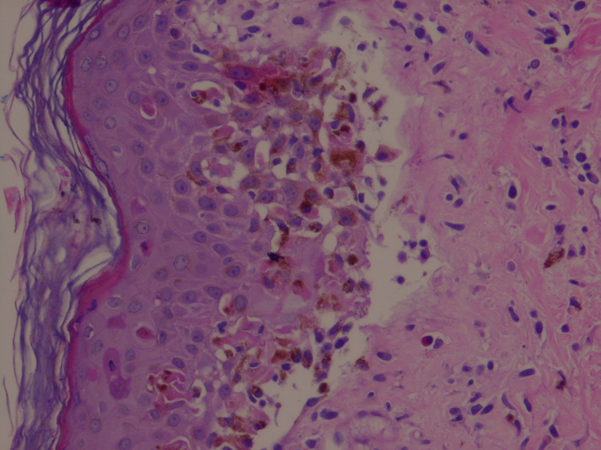
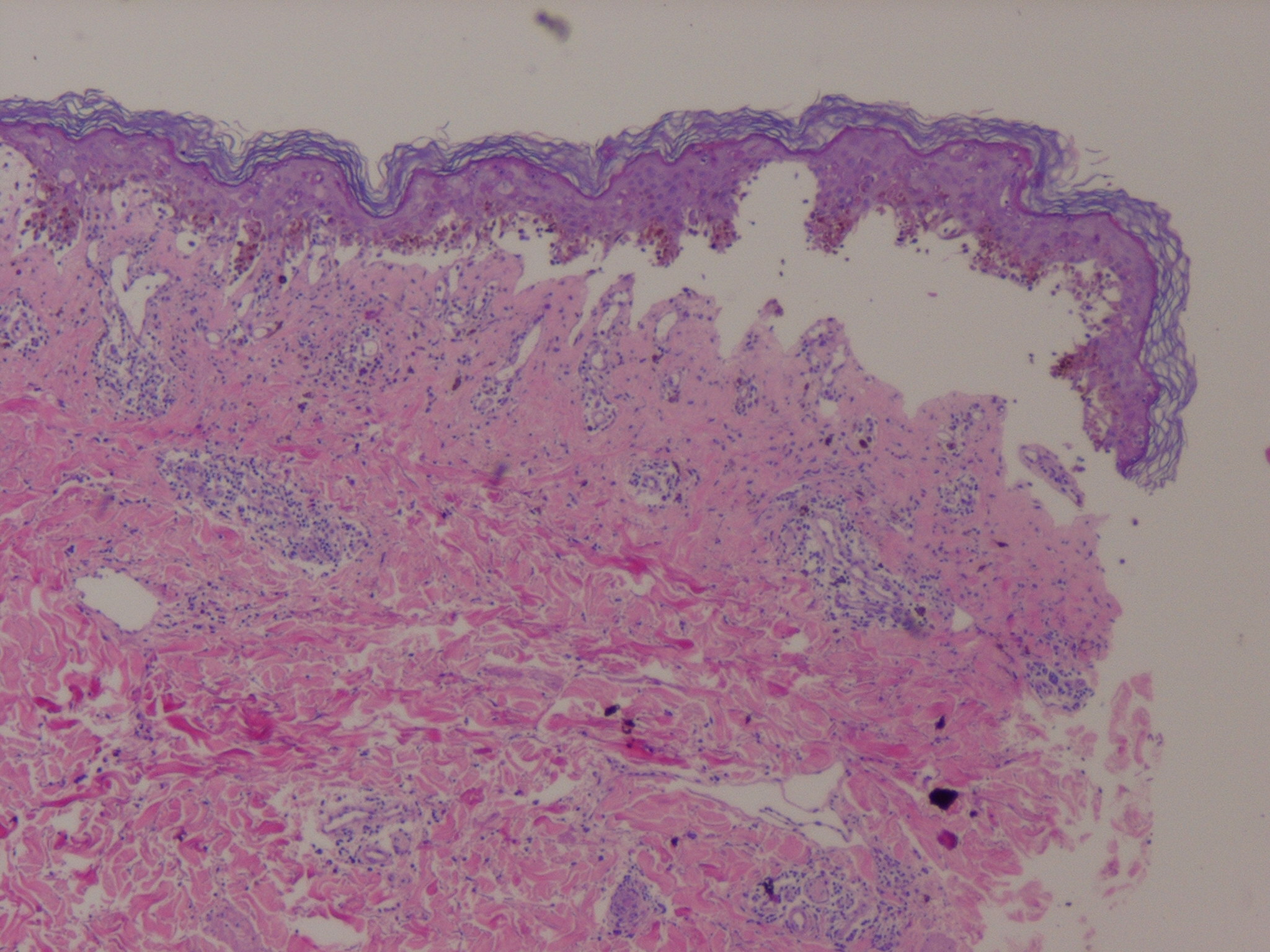
Related Physical Findings Fever and other constitutional symptoms are usually absent in EM minor, and the physical examination is normal. Fever higher than 38.5°C (101.3°F) is present in 32 percent of cases of EMM. Mouth erosions may be very painful and may impair alimentation. The patient may be unable to close the mouth and may constantly drool blood-stained saliva. Cervical lymphadenopathy is usually present in these patients. The pain of genital erosions may lead to reflex urinary retention. Cough, polypnea, and hypoxia may occur in M. pneumoniae-related cases. LABORATORY FINDINGS Histopathologic Analysis Early lesions of EM exhibit lymphocyte accumulation at the dermal-epidermal interface, with exocytosis into the epidermis, lymphocytes attached to scattered necrotic keratinocytes (satellite cell necrosis), spongiosis, vacuolar degeneration of the basal cell layer, and focal junctional and sub-epidermal cleft formation . The papillary dermis may be edematous but principally contains a dense mononuclear cell infiltrate, which is more abundant in older lesions. The vessels are ectatic with swollen endothelial cells; there may be extravasated erythrocytes and eosinophils. Immunofluorescence findings are negative or non-specific. In advanced lesions sub-epidermal blister formation may occur, but necrosis rarely involves the entire epidermis . In late lesions, melanophages may be prominent. The histopathologic appearance of EM lesions is different from that of SJS-TEN lesions, in which dermal inflammation is moderate to absent and epidermal necrosis much more pronounced . Still, the histopathologic appearances are somewhat overlapping and do not allow the distinction of EM from SJS-TEN in all instances. The main reason for performing a biopsy is to rule out other diagnoses. Other Laboratory Tests There are no specific laboratory tests for EM. In more severe cases, an elevated erythrocyte sedimentation rate, moderate leukocytosis, increased levels of acute-phase proteins, and mildly elevated liver aminotransferase levels may occur. In the presence of respiratory symptoms a chest radiograph is needed, and documentation of M. pneumoniae infection by PCR assay of a throat swab and serologic testing (a pair at a 2- or 3-week interval) should be sought. Investigations to document causality are important in cases with frequent recurrences when prevention with long-term antiviral treatment is considered and when there is no clinical evidence of association with herpes. HSV can sometimes be isolated from the initial lesion of labial herpes. Amplification of HSV Pol gene from biopsy samples of EM lesions is not done routinely. A negative result on serologic testing for HSV may be helpful to exclude the possibility of herpes-associated EM. The positive predictive value of the presence of HLA-DQB1*0301 is too low to have any clinical value. In a retrospective analysis of 66 pediatric cases discharged from hospital with a diagnosis of EM 24 (36 percent) were clearly not EM or SJS.7 Diseases that had been frequently erroneously called EM were urticaria and maculopapular drug eruption. The designation of Rowell syndrome11 is used for a variety of cutaneous lupus erythematosus with lesions resembling those of EM. Subacute evolution, a positive result on direct fluorescence testing, and the presence of anti-nuclear antibodies exclude EM. Sweet syndrome can mimic EM minor; biopsy easily distinguishes the two. Paraneoplastic pemphigus and more rarely other autoimmune blistering diseases occasionally present with target-like lesions that can be confused with those of EM. One of these has been reported as EMM with antidesmoplakin antibodies.12 It resembles EMM in its acute and recurrent course, but the presence of acantholysis, deposits of immunoglobulin G around basal cells, and serum antibodies against desmoplakin distinguish it from EM. Whether considered a variant of EMM or a separate disease, SJS should be recognized promptly for three reasons: (1) the possibility of life-threatening complications, (2) the risk of progression to TEN, and (3) the need for urgent withdrawal of suspected causative drug(s). Pain, constitutional symptoms,
▪ COURSE AND COMPLICATIONS EM runs a mild course in most cases, and each individual attack subsides within 1 to 4 weeks. Recovery is complete, and there are usually no sequelae, except for transient hypopigmentation or hyperpigmentation in some cases. In rare instances the ocular erosions of EMM may cause severe residual scarring of the eye. M. pneumoniae-related EMM may be associated with severe erosive bronchitis. Differential Diagnosis of Erythema Multiforme (EM)
Recurrences are common and may characterize the majority of cases. In one report of a large series of patients with recurrent EM, the mean number of attacks was 6 per year (range, 2 to 36), and the mean total duration of disease was 9.5 years. In 33 percent, the condition persisted for more than 10 years.13 Up to 50 recurrences have been described in a single patient. The severity of episodes in patients with recurrent EM is highly variable and unpredictable. The frequency of episodes and cumulative duration of disease are not correlated with the severity of attacks. The frequency and severity of recurrent EM tend to decrease spontaneously over time (after 2 years or longer), parallel with the improvement of recurring HSV infection. A small fraction of patients experience a prolonged series of overlapping attacks of EM; this has been labeled continuous EM or persistent EM. TREATMENT The aims of treatment are to reduce the duration of fever, eruption, and hospitalization. Based on retrospective series or small controlled trials, the use of systemic corticosteroids seems to shorten the duration of fever and eruption but may increase the length of hospitalization because of complications. However,
Several series indicate that administering anti-HSV drugs for the treatment of full-blown postherpetic EM is useless. When symptomatic, M. pneumoniae infection should be treated with antibiotics (macrolides in children, macrolides or quinolone in adults). There is no evidence indicating whether it improves the evolution of the associated EM. Therefore, when asymptomatic infection is suggested by serologic testing, treatment is not mandatory. Liquid antacids, topical glucocorticoids, and local anesthetics relieve symptoms of painful mouth erosions. PREVENTION Continuous therapy with oral anti-HSV drugs is effective to prevent recurrences of herpes-associated EM with or without clinical evidence that herpes is the precipitating factor.6 Topical acyclovir therapy used in a prophylactic manner does not prevent recurrent herpetic EM. In a series of 65 patients with recurrent EM, 11 were treated with azathioprine when all other treatments had failed. Azathioprine was beneficial in all 11 patients. Retrospective uncontrolled analyses of thalidomide therapy have indicated that it is moderately effective for the treatment of EM. In one randomized controlled trial, levamisole appeared useful. Because agranulocytosis is a severe and not exceptional adverse effect, levamisole use is permitted in only a few countries. The benefit-risk ratio is probably too low to support its use in the treatment of EM.
|
||||||||||||||||||||||||||||||||||||||||||||||||||||||||||