|
Alopecia mucinosa = الحاصة المخاطينية |
 |
 |
 |
|
Alopecia Mucinosa
Follicular mucinosis is characterized clinically by grouped erythematous papules and/or plaques that may be markedly indurated or nodular ( and histologically by mucin accumulation in hair follicles . It can be classified into two types: a primary (idiopathic) type and a secondary variety. The primary form tends to have a shorter but benign course. The secondary type has been associated with numerous benign and malignant conditions, including lymphomas, of which the majority are mycosis fungoides. A distinct variant of mycosis
|
|
fungoides-follicular mycosis fungoides-may or may not be associated with follicular mucinosis .
|
|
The primary form tends to affect children and young adults more frequently and resolves spontaneously in several months (acute benign type) or several years (chronic benign type) . It is often confined to the head and neck but may be disseminated. The secondary type tends to form more widespread plaques and is almost always a disorder of adults.
|
|
The secondary type has been found in association with other Iymphoproliferative disorders, including Hodgkin's disease , cutaneous B-celllymphoma , acute myeloblastic leukemia , chronic lymphocytic leukemia , and syringolymphoid hyperplasia with cutaneous T-cell lymphoma , as well as a number of inflammatory cutaneous disorders such as chronic discoid lupus erythematosus , angiolymphoid hyperplasia , alopecia areata (, eosinophilic pustular folliculitis , spongiotic dermatitis, lichen striatus, arthropod bites, sarcoidosis, Goodpasture's syndrome , leprosy , and growths such as verrucae (, melanocytic nevi, and squamous cell carcinoma of the tongue . The numerous associations with production of follicular mucin certainly suggest that this is a relatively nonspecific reaction pattern.
|
|
There has been controversy about whether the histopathology allows for distinction between the primary and secondary forms. Initially it was considered that the histopathology could be predictive (. Later, some researchers claimed that transition from the benign form to a lymphomatous type could occur , whereas others disputed this finding . In 1989 a study of 59 cases concluded that there was no clinical or pathologic pattem by which the ultimate outcome of the condition could be predicted .
Others have reported that adults older than the age of 40 years with widespread follicular mucinosis are at increased risk for mycosis fungoides or Sezary's syndrome , although Cerroni et al. claimed that criteria purported to differentiate lymphoma-associated follicular mucinosis from the idiopathic type were not effective . A long-term follow-up study (median 10 years) of seven patients younger than 40 years of with primary follicular mucinosis failed to demonstrate progression to cutaneous T-cell lymphoma, despite the presence of a Tcell clone in five of the patients .
|
|
In 1957, Pinkus described alopecia mucinosa, the term used when follicular mucinosis affects terminal hairbearing areas and is associated with hair loss . Papules and plaques may be present or inconspicuous in this form, which may show only alopecia. Scarring is seen more commonly when alopecia mucinosa is associated with cutaneous T-cell lymphoma. Others claim that alopecia mucinosa is simply one of the many morphologic variants of mycosis fungoides , whereas LeBoit and LeBoit acknowledged the paradoxes of alopecia mucinosa that have led to the debate over classification (.
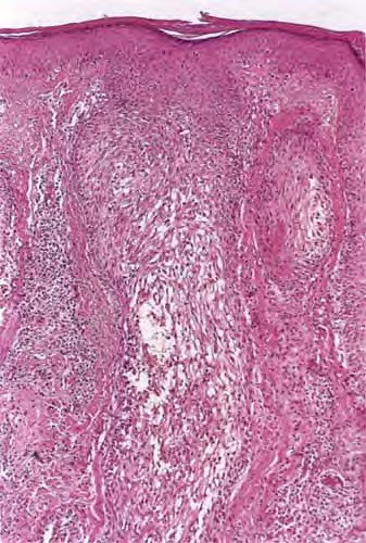
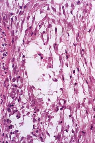
Histopathology.
Within the outer root sheath and sebaceous gland epithelium, there is reticular epithelial degeneration that sometimes evolves into more extensive cavitation within which mucin is deposited . Occasionally, little mucin can be detected, perhaps because of removal of this water-soluble material in the processing procedure. The deposited mucin is an acid mucopolysaccharide that stains metachromatically with toluidine blue at pH 3.0, as well as with Alcian blue at acid pH. The fact that it can be substantially removed by digestion with hyaluronidase demonstrates that the mucin is predominantly hyaluronic acid. Colloidal iron stain may also be used for its detection.
|
|
Inflammation is composed of lymphocytes and histiocytes, but there can also be eosinophils. There may be exocytosis into the outer root sheath epithelium of the infundibulum and the sebaceous gland epithelium. Although individual pathologic criteria are not absolutely diagnostic of the type of
|
|
follicular mucinosis (primary or secondary), features that have been proposed as favoring a lymphoma-associated lesion include an atypical lymphocytic infiltrate or increased density of the perifollicular infiltrate with substantial folliculotropism . This study also suggested that a prominent eosinophilic infiltrate and more substantial mucin deposition tend to favor a benign process, but a subsequent study failed to substantiate these findings .
|
|
Pathogenesis.
Electron microscopic studies have shown that the mucin is a product of the outer root sheath epithelial cells. The cytoplasm shows prominent, dilated, rough-surfaced endoplasmic reticulum containing fine, granular, filamentous material that is secreted into the intercellular spaces . In two of our patients with primary, idiopathic alopecia mucinosa with reversible alopecia, we found, using transverse sections, an increased
|
|
number of resting-phase follicles, mostly catagen, ranging from 46% to 92% of all terminal follicles (unpublished observation).
|
|
|
|
|