| Hydroa vacciniforme = المائية اللقاحينية |
|
|
Hydroa Vacciniforme
EPIDEMIOLOGY
Hydroa vacciniforme (HV) occurs in the United States, the United Kingdom, continental Europe, Japan, and very possibly elsewhere. However, its rarity and lack of universally acknowledged diagnostic criteria make precise evaluation difficult.
ETIOLOGY AND PATHOGENESIS
The pathogenesis of HV is unknown. No chromophores have been identified, and although the UVB minimal erythemal dose reaction is normal in most patients, some have reduced UVA values. Blood, urine, and stool porphyrin concentrations are normal, as are all other laboratory parameters. Nevertheless, the relationship of the eruption to sunlight exposure, its distribution, and its early clinical appearances are all very similar to those of PMLE, which strongly suggests a possible relationship to that disorder. On the other hand, the fully developed HV eruption is much more severe than that of PMLE, is always associated with permanent pock scarring, and is unresponsive to treatments effective for PMLE, apart
HYDROA VACCINIFORME AT AGLANCE
CLINICAL FINDINGS
History.
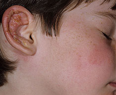
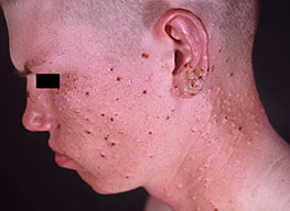
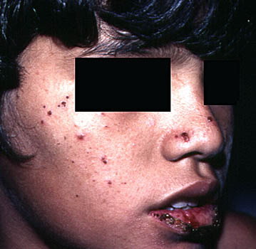
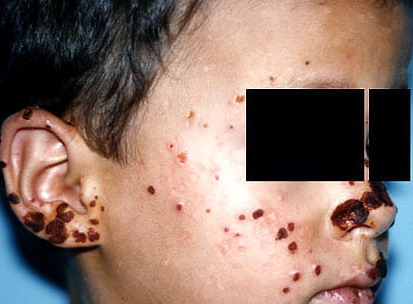
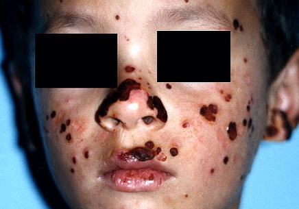
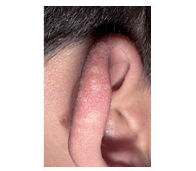
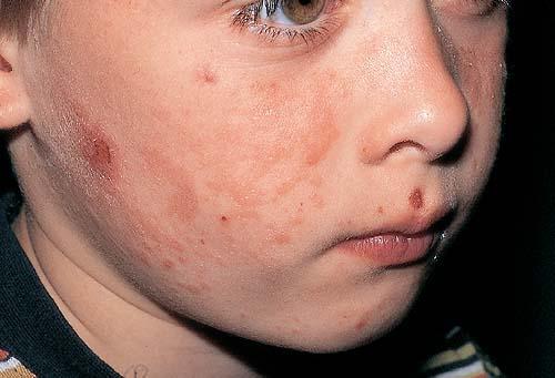
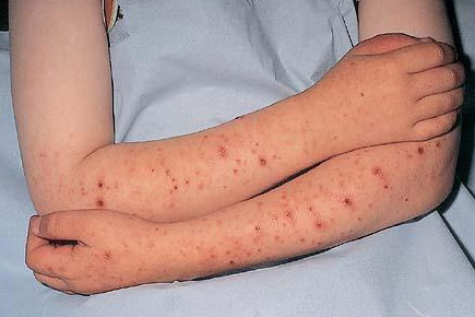
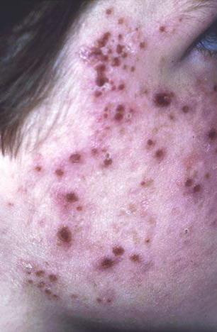
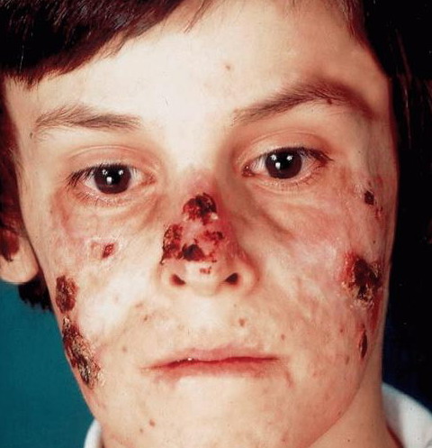
Most HV arises in early childhood and resolves spontaneously by puberty, although some patients suffer lifelong. There is a male predominance for severe forms, whereas milder disease is more common in females. Familial incidence is exceptional. One estimate of the prevalence of HV is 0.34 cases per 100,000 with an approximately equal sex ratio; males had a later onset and longer duration of the disorder than females. The HV eruption typically occurs in summer, with an often intense burning or stinging sensation followed by the appearance of individual or confluent papules and then vesicles within hours of sun exposure. This is followed by their umbilication, crusting, and progression to permanent pock scarring within weeks. The rash affects the cheeks and to a lesser extent other areas of the face, as well as usually the backs of the hands and outer aspects of the arms, generally symmetrically. It also rarely affects other sites.
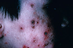
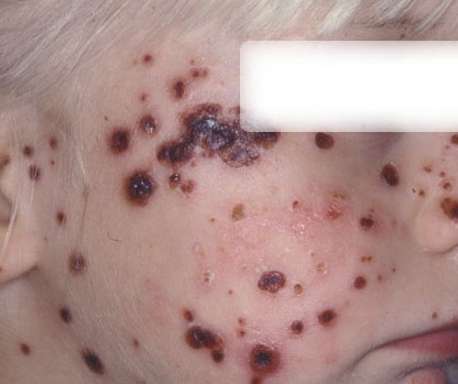
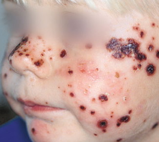
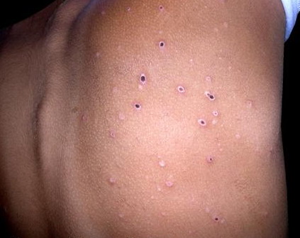
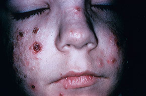
Cutaneous Lesions.
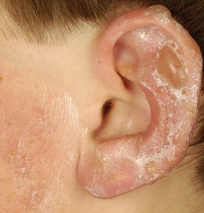
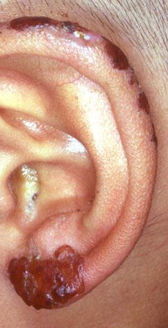
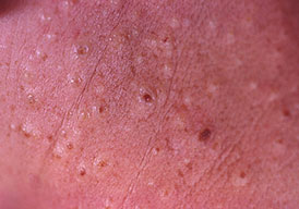
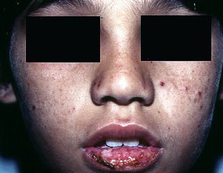
HV is characterized by initial erythema, sometimes with swelling, followed by symmetrically scattered, usually tender papules within 24 hours; vesiculation, occasionally confluent and hemorrhagic (Fig. 90-6); umbilication; then crusting and detachment of the lesions with permanent, depressed, hypopigmented scars within weeks. Oral ulcers and eye signs also rarely occur.49,50
LABORATORY TESTS
Histology.
Early histologic changes include intraepidermal vesicle formation with later focal epidermal keratinocyte necrosis and spongiosis in association with dermal perivascular neutrophil and lymphocyte infiltration. Older lesions show necrosis, ulceration, and scarring. Vasculitic features have been reported in rare cases.
Blood Tests.
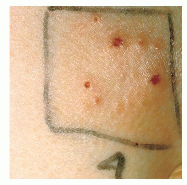
Blood, urine, and stool porphyrin concentrations should be assessed to exclude cutaneous porphyria, as should the circulating anti-nuclear factor and extractable nuclear antibody titers to exclude the slight possibility of cutaneous lupus. Phototesting. Phototesting may show reduced minimal erythema doses in short-wavelength UVA in some cases but is not usually able to discriminate from other photodermatoses. Simulated solar irradiation may also induce erythema at reduced irradiation doses or occasionally provoke the typical vesiculation of HV . Other Tests. Viral studies to check for herpes infection or other viral disorders should be undertaken if photo-exacerbation or photo-induction of such a disorder seems at all possible.
COMPLICATIONS Pock scarring is an inevitable accompaniment of HV.
PROGNOSIS AND CLINICAL COURSE HV often resolves in adolescence but may occasionally persist into adult life.
PREVENTION Sun avoidance and sunscreen use, as well as prophylactic phototherapy annually in spring, tend to prevent HV in some patients. TREATMENT The treatment of HV consists of restriction of sun exposure and use of high-protection-factor broad-spectrum sunscreens. Occasionally, antimalarials appear to have helped, but their true value is not established. Similar observations have been made for β-carotene, which in our hands was ineffective in three cases. As with PMLE, prophylactic phototherapy with narrowband UVB or
Box 90-3 Differential Diagnosis of Hydroa Vacciniforme
|