
Tinea Favosa
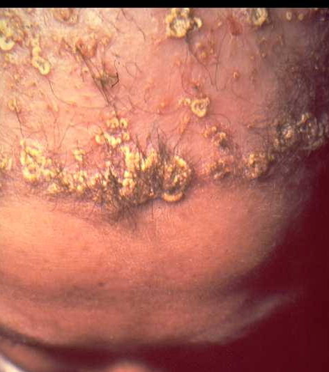
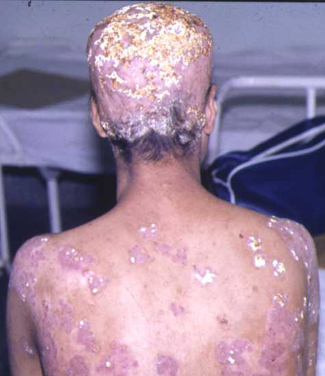
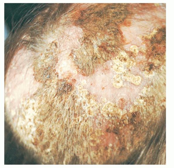
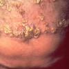
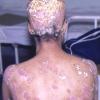
Tinea favosa or favus (Latin, “honeycomb”) is a chronic dermatophyte infection of the scalp, glabrous skin, and/or nails characterized by thick yellow
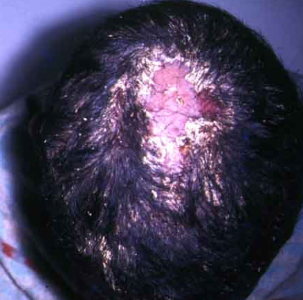
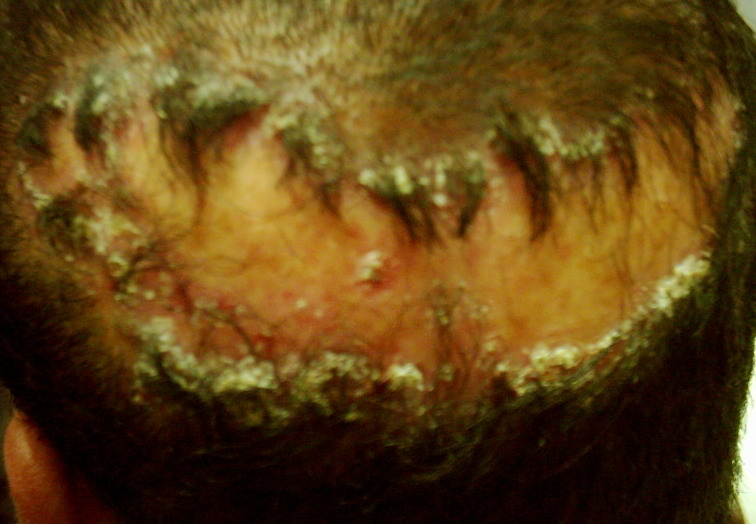
crusts (scutula) within the hair follicles, which lead to scarring alopecia.
Differential Diagnosis of Tinea Capitis
Most Likely
· Seborrheic dermatitis, atopic dermatitis, impetigo and pustular or plaque psoriasis, bacterial pyodermas, folliculitis decalvans, and perifolliculitis capitis abscedens et suffodiens
Consider
· Alopecia areata, trichotillomania, pseudopelade
Rule Out
· Syphilis, lupus erythematosus
EPIDEMIOLOGY
Favus is usually acquired before adolescence and extends into adulthood. Associated with malnutrition and poor hygiene, it has become geographically limited in the past century, as it is now seen almost exclusively in Africa, the Middle East, and parts of South America. Even in these regions, its incidence is dramatically declining.
ETIOLOGY
T. schoenleinii is the most common cause of human favus, with T. violaceum and M. gypseum occurring as rare isolates. Although favus occurs in poultry, mice, and horses, there are very few reports in the literature of human favus caused by an organism responsible for animal favus.
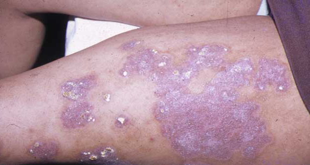
CLINICAL FINDINGS
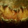
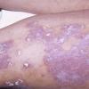
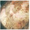
Early favus (usually the first 3 weeks of infection) is characterized by patchy follicular erythema with slight perifollicular scaling and mild matting of the hair. Progressive hyphal invasion distends the follicle, first producing a yellowish-red papule, then a yellow concave crust often centered about a single dull, dry hair that is less brittle than that of endothrix infections. The scutulum may reach 1 cm in diameter, engulfing surrounding hairs and coalescing with other scutula to form large adherent mats with an unpleasant cheese-like or musky odor. Over several years, the lesions advance peripherally, leaving central, atrophic clearings of alopecia.
LABORATORY TESTS
T. schoenleinii exhibits subtle, grayish-green fluorescence along the entire hair with Wood's lamp examination . Microscopy with KOH preparation reveals hyphae arranged lengthwise around and within the hair shaft, rare arthroconidia, and vacant air spaces.