|
Dermatologic Manifestations of Enteroviral Infections
The human enterovirus genus is a member of the Picornaviridae family of small, icosahedral, single-stranded, positive-sense RNA viruses. A total of 92 serotypes are currently recognized by the International Committee on Taxonomy of Viruses classification1 :
- Human enterovirus type A (17 serotypes): These include coxsackie A virus types 2-10, 12, 14, and 16 and echovirus type 71. New enterovirus serotypes were described in 2005 (types 76, 89, 90, and 91), and it is believed that they are a subgroup of human enterovirus type A.2
- Human enterovirus type B (56 serotypes): These include coxsackie A virus type 9; coxsackie B virus types 1-6; echovirus types 1-7, 9, 11-21, 24-27, and 29-33; and enterovirus type 69. The 13 new serotypes reported in 2007 (enterovirus types 79-88, 97, 100, and 101) are probably members of human enterovirus type B.2
- Human enterovirus type C (16 serotypes) - The most well known are coxsackie A virus types 1, 11, 13, 15, 17-22, and 24. Poliovirus types 1-3 have been included in this group.
- Human enterovirus type D - Enterovirus types 68 and 70, including several newly identified serotypes, such as enterovirus 73-753,4 and enterovirus types 77-785
Human enteroviruses are distributed worldwide, with 2 major patterns of infections, endemic and epidemic, within a given geographical area.1
Enteroviruses usually cause transient, often subclinical, infections. Enteroviruses are also responsible for a wide variety of syndromes, including exanthematic fever, enteritis, encephalitis, aseptic meningitis, myocarditis, and respiratory tract infections. Coxsackieviruses, echoviruses, and enterovirus type 71 are also significant causes of cutaneous disease. A relationship between enterovirus RNA and chronic fatigue syndrome has been described,6 and these viruses could initiate and perpetuate the immunological response seen in chronic fatigue syndrome.
Coxsackieviruses are separated into 2 groups, A and B. Coxsackie A viruses are the primary etiologic agents of herpangina and hand-foot-and-mouth disease (HFMD). Coxsackie B viruses are associated with epidemic pleurodynia (ie, Bornholm disease), epidemic myalgia, myocarditis, and pericarditis. Bowles et al suggest that coxsackie B virus may be an etiologic agent of juvenile dermatomyositis.7
Zahorsky first described the clinical spectrum of herpangina in 1920. Later, coxsackie A virus was isolated from pharyngeal washings and stool samples of patients with herpangina. Subsequently, many reports have confirmed this association. Robinson et al first isolated the coxsackie A virus type 16 serotype in 1957 during a Canadian epidemic of exanthema and stomatitis.8 Two years later, Alsop et al used the term hand-foot-and-mouth disease to describe a similar eruption in England.9
Echoviruses include 34 distinct serotypes, and at least half can cause a rash. The 2 skin diseases specifically associated with echoviruses are Boston exanthem disease (BED), caused by echovirus type 16, and eruptive pseudoangiomatosis, (EP) caused by echovirus types 25 and 32.
Pathophysiology
Enteroviruses are spread from person to person by oral-oral and fecal-oral routes and, because the virus can be isolated from cutaneous and ocular lesions, presumably, may also be transmitted through direct contact with fluid from these cutaneous and ocular lesions.
Another source of infection could be swimming pools because enteroviruses are easily detectable in natural and treated water sources.
It has been suggested that enteroviruses can be transmitted antenatally, either transplacentally or potentially via ascending infection. When maternal enterovirus infection is acquired during late pregnancy, vertical transmission has been shown to be relatively common.2
The viral incubation period is usually 2-5 days. The viruses are highly contagious, and they are a common cause of widespread outbreaks. After the ingestion of infectious material, enteroviruses are implanted and replicated in the alimentary tract (nasopharynx and ileum). If local replication is limited, the disease remains asymptomatic. If the virus passes into the regional lymphatic nodes and the reticuloendothelial system organs, minor or nonspecific disease may develop. Virus may also spread by the hematologic route, which results in a more severe and characteristically systemic illness.
Immune activation by the enterovirus leads to the production of immunoglobulin M (IgM) type-specific antibodies, which may be detected in the serum 1 week after infection. They are responsible for neutralization and rapid elimination of the virus from the blood and other sites of implantation. Serum IgM antibodies can be detected for 6 months after the patient's recovery, and convalescent immunoglobulin G can be detected for 1-2 years. Most enteroviral infections confer lifelong immunity to the serotype-specific agent. In addition, antibodies to these related viruses are known to cross-react, and they do so in different patterns, based on the country, serotype, and specific population, making comparisons of disease-based studies amongst these groups difficult.
History
Coxsackieviruses and echoviruses cause many nonspecific exanthems and enanthems. An exanthem (ie, nonspecific morbilliform eruption) normally occurs 3-4 days before the characteristic enanthem (ie, oral vesicles) appears
Seasonal distribution is a characteristic feature. In temperate climates, enteroviral infections are most common in the summer and autumn, whereas in tropical areas, they tend to occur year round. Some of the more specific clinical syndromes are described below.
- Herpangina
- The principle cause is coxsackie A viruses (serotypes 2-6, 8, and 10). Other viral etiologies include coxsackie B viruses (serotypes 1-4), echoviruses, adenoviruses, and other enteroviruses.
- After an incubation period of approximately 4 days, the disease begins with acute onset of fever (temperature range, 38.5-40°C) accompanied by headache, sore throat, dysphagia, anorexia, and, occasionally, vomiting and abdominal pain.
- Hand-foot-and-mouth disease
- HFMD is most commonly associated with coxsackie A virus 16. HFMD is also associated with infection by coxsackie A virus serotypes 4-7, 9, and 10; coxsackie B virus serotypes 2 and 5; and enterovirus 71. The incubation time is 1-7 days.
- A brief prodromal period is characterized by low-grade fever, malaise, abdominal pain, and/or respiratory symptoms.
- Prominent historical features include oral pain and odynophagia, painless vesicles on the hands and feet, and a morbilliform eruption on the buttocks.
- Boston exanthem disease
- BED is caused by echovirus 16.
- Similar to HFMD, BED begins with a brief febrile prodrome.
- Eruptive pseudoangiomatosis11
- EP is associated with echovirus 25 and echovirus 32.
- An initial viral prodrome is characteristic.
Physical
- Herpangina
- The enanthem is characterized by the presence of gray-white minute papulovesicles approximately 1-2 mm in diameter.
- The lesions are surrounded by an erythematous halo, which progresses to a shallow ulcer covered by fibrin.
- The lesions are self-limiting, resolving over 5-10 days.
- Lesions are most frequently found on tonsils, uvula, soft palate, and anterior pillars of the tonsillar fauces.
- The most important differential diagnosis to be considered is acute herpetic gingivostomatitis. However, acute gingivitis is not present in herpangina. Furthermore, herpetic gingivostomatitis is characterized by longer duration and more severe pain.
- Hand-foot-and-mouth disease
- Oral lesions begin as erythematous macules and papules that are 2-8 mm in diameter; these progress to form thin-walled vesicles. The vesicles rapidly ulcerate, remaining as shallow painful ulcers surrounded by an erythematous halo. Lesions heal without treatment over 5-10 days. The lesions may be found anywhere in the oral cavity, but they most frequently appear on the hard palate, tongue, buccal mucosa, and gums. The tongue may be erythematous and edematous, and pain may interfere with adequate oral intake.
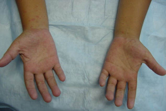
- Skin lesions are variably present, but they are characteristic when they occur. The lesions appear along with or shortly after the oral lesions appear, and they may vary in number from a few to more than 100. They begin as erythematous macules or papules, which quickly become small (as large as 5 mm in diameter), gray, oval or linear vesicles surrounded by a red halo. The hands are more commonly involved than the feet. Lesions usually occur on the lateral aspects of the fingers and toes, especially around the nails, but they may be seen in the digital flexures and on the palms and soles. The lesions gradually disappear over 7-10 days, without scarring.
- In some patients, especially infants, a more widespread papular or vesicular exanthem appears principally on the buttocks, although it may occasionally generalize.
- In Asia, some epidemics of HFMD have been associated with severe refractory left ventricular failure, cardiogenic shock, CNS disorders, and death. These cases have generally been linked to enterovirus 71.
- Boston exanthem disease
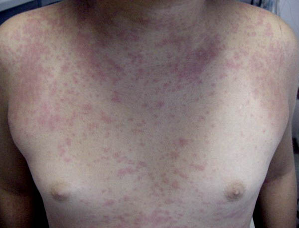
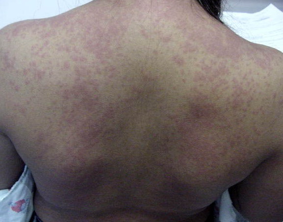
- After a brief febrile illness, pink macules and papules abruptly erupt on the face, trunk, and, less commonly, on the extremities.
- Small ulcerations may be seen on the soft palate and tonsils.
- Eruptive pseudoangiomatosis
- After the prodromal period, 2- to 4-mm blanchable, red papules resembling cherry angiomas appear.
- The lesions usually number no more than 10, and they resolve spontaneously within 10 days.
- They are distributed on the face, trunk, and extremities.
- Other associations
- Acute hemorrhagic conjunctivitis12,13,4 is linked to enterovirus 70 and coxsackie A virus 24.
- Pustular stomatitis associated with erythema multiforme is linked to coxsackie B virus 5.14
- Widespread vesicular eruption is linked to coxsackie A virus 4.
- Gianotti-Crosti–like eruption is linked to coxsackie A virus 16.
- Rubelliform eruption is linked to echovirus 2.
- Morbilliform eruption is linked to echoviruses 6, 11, and 25.
- Rubelliform or morbilliform eruption is linked to echovirus 9.
- Petechiae are linked to echoviruses 11 and 19.
- Punctate macular eruption is linked to echovirus 19.
- Vesicular eruption is linked to echovirus 11.
- Some epidemiological studies strongly suggest that coxsackievirus infections, in particular type B coxsackieviruses, are related to the induction or exacerbation of type 1 diabetes.
|

