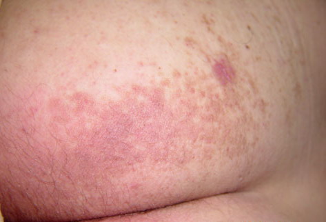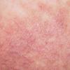
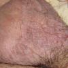
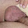
Darier like epidermal naevus
Darier disease in a unilateral or localized pattern was first reported at the turn of the last century. This variant often lacked other features that were associated with typical Darier disease and the skin lesions were usually confined to a limited area [1].
The lack of a family history and absence of other signs of DD, as well as the limited distribution of the lesions in many patients, are in favor of a nevoid origin. Starink and Woerdeman reported seven cases showing unilateral, linear, or zosteriform patterns, without other findings of DD and suggested the name acantholytic dyskeratotic epidermal nevus [2]. In a review of 167 biopsy specimens taken from patients with epidermal nevi, Su observed only two specimens (1.2%) with DD-like changes [3].
A singular report described localized DD affecting the scalp of a 5-year-old girl and of her mother, without other cutaneous abnormalities, possibly suggesting an autosomal dominant inheritance pattern [4]. Two other reports have associated typical localized DD with palmo-plantar papules and nail disorders characteristic of DD [5, 6].
The abnormal keratinocyte adhesion of DD is inherited in an autosomal dominant pattern, and the causative gene is located at chromosome 12, 12q23-24.1 [7]. The linear form of Darier disease could result from genetic mosaicism for this autosomal dominant disorder. This mutation occurs during embryogenesis [7, 8]. Two types of segmental mosaicism have been described recently. In the first type (type 1), the cutaneous manifestations show the same degree of severity that is found in the corresponding non-mosaic trait. In the second type (type 2), the segmentally involved lesions are much more severe and may be superimposed on a milder diffuse form of the same disease [8].
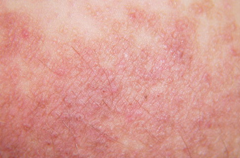
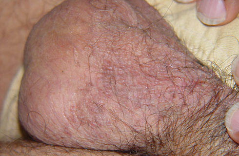
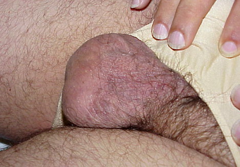
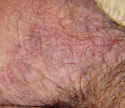
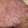
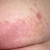
Our patient had a more pronounced involvement than the usual Darier disease lesions, disposed in a linear arrangement, with nail features. He probably presents a type 2 segmental manifestation of the disorder.