| Bowenoid papulosis= الداء الحطاطي البوفناني |
|
|
Bowenoid Papulosis
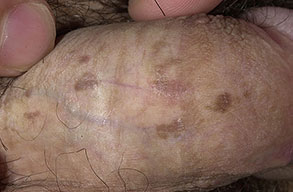
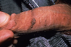
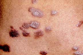
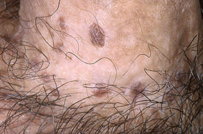
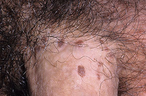
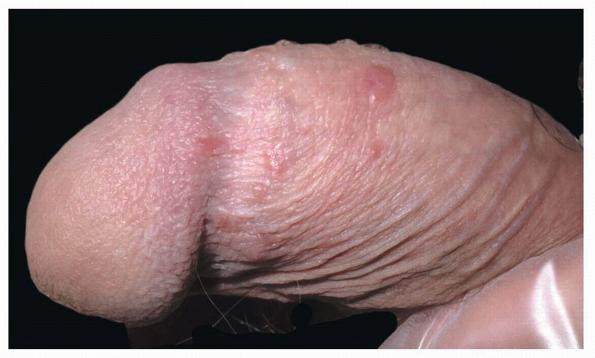
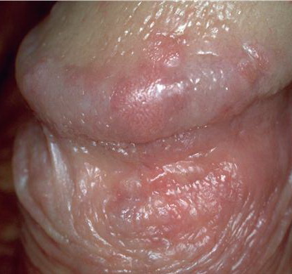
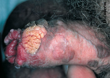
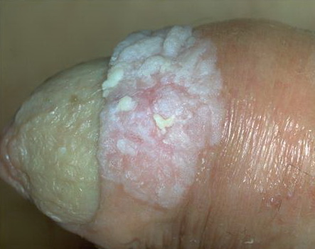
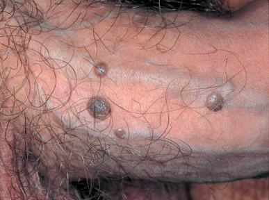
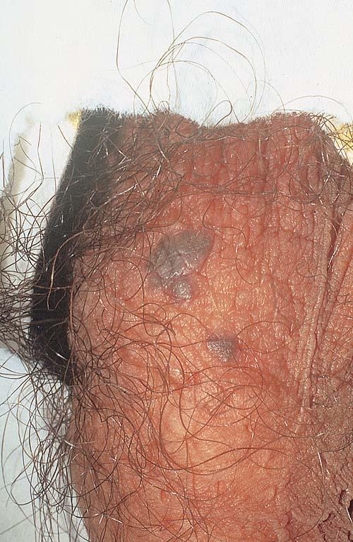
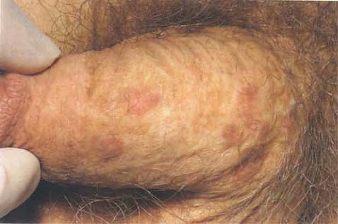
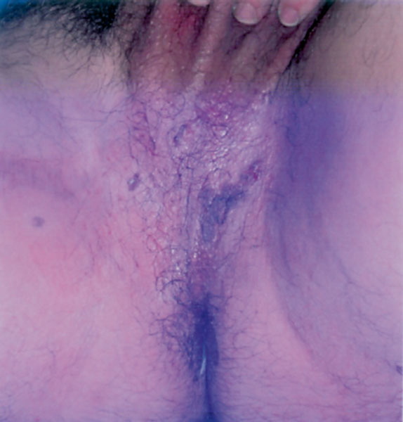
Bowenoid papulosis (BP) is characterized clinically by the presence of pigmented verrucous papules and plaques primarily on the genitalia of predisposed individuals, and histopathologically by the presence of SCC in situ-like changes . BP is caused by infection with HPV, and numerous HPV types have been linked to BP, including types 42, 48, and 51 to 54 . Lesions are pink, reddish brown, or violaceous Differential diagnosis includes early condylomata acuminata Histopathologically, the epidermis is usually hyperplastic with atypia, disordered maturation, scattered mitotic figures, and dyskeratotic keratinocytes. The course of BP is variable, ranging from spontaneous regression to persistence of lesions to transformation into BD and invasive SCC. Patients with BP and their sexual partners should be followed and examined periodically, because of the risk of developing SCC, cervical, and vulvar neoplasia. Patients with persistent disease should probably undergo testing for altered immune status. Treatment of BP is recommended, and it typically responds well to local therapy, although recurrences are common. Therapeutic options include local destructive measures such as curettage
VIRAL KERATOSES AT A GLANCE
· Two viral keratoses with the potential for malignant transformation are bowenoid papulosis (BP) and epidermodysplasia verruciformis (EV). · BP is a precancerous condition of the genitalia caused by infection with human papillomavirus (HPV); oncogenic types 16, 18, and 33 are most commonly involved. · BP may regress spontaneously, persist, or transform into Bowen disease or squamous cell carcinoma (SCC). · Treatment options include topical imiquimod, curettage, excision, and laser vaporization. · Sexual partners of patients must be examined and followed closely for development of cervical, vulvar, or penile carcinoma. · EV is a rare autosomal recessive disease associated with mutations in the EVER1 and EVER2 genes. · Affected individuals show a propensity toward infection with certain strains of HPV-5 and HPV-8. · Clinical presentation is widespread flat wart-like papules and plaques in childhood; risk of developing SCC later in life is high. · Sun avoidance, sun-protective measures, regular dermatologic follow-up, and screening of family members for the disease is important.
Epidermodysplasia Verruciformis
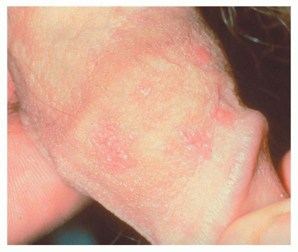
Epidermodysplasia verruciformis (EV) is a rare autosomal recessive genetic disease that manifests in childhood with disseminated flat, wart-like papules and plaques. These individuals are at high risk for the development of SCC in these wart-like lesions, which result from an abnormal propensity to infection with certain HPV sub-types, most notably HPV-5 and HPV-8 . A susceptibility locus for EV was localized to chromosome band 17q25 in 1999, and mutations in two novel genes from this same region (EVER1 and EVER2) were identified and associated with EV more recently. These patients present with numerous thin, pink, flat papules and plaques that resemble verruca plana. They also may have widespread scaly, erythematous, or hypopigmented macules and flat papules that appear similar to tinea versicolor
▪ SQUAMOUS CELL CARCINOMA IN SITU (BOWEN DISEASE)
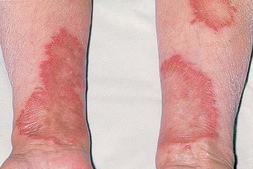
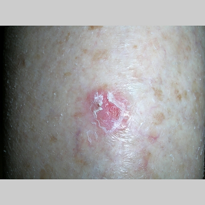
BD is SCC in situ, originally described in 1912 by John T. Bowen, a Boston dermatologist.114 It affects both skin and mucous membranes and has the potential to progress to invasive SCC. Epidemiology BD may occur at any age in adults, but it is rarely seen in individuals younger than 30 years. The typical patient with BD is older than 60 years. The disease is said to occur with an equal incidence in men and women, although most studies report a slight preponderance in women. BD can be found on any body site, including both sun-exposed and non-sun-exposed regions of the body, although it appears to have a predilection for sun-exposed surfaces such as the head and neck and for the lower legs of women, in particular. The exact incidence of BD in the United States is unknown, but in one population study in Hawaii, the incidence was estimated at 142 per 100,000 persons.116 Lesions of BD are usually solitary but are multiple in up to 10 percent to 20 percent of individuals. Etiology and Pathogenesis A number of different factors have been implicated in the development of BD, including a history of significant sun exposure, arsenic exposure, ionizing radiation, immunosuppression, and certain types of HPV infection. Up to 30 percent of extragenital BD lesions have been found to harbor HPV DNA. The age group and sites of predilection of BD suggest a strong association with sun exposure. BD is also rare in more heavily pigmented individuals, and it has been described with increased frequency in patients undergoing PUVA therapy. The association with arsenic exposure has already been discussed. SCC in situ is seen commonly in organ transplant recipients after years of immunosuppressive drug therapy. Infection with HPV has been implicated in causing certain sub-types of BD. In particular, HPV-16 has been detected in many cases of anogenital BD and in some cases of finger and periungual BD. Clinical Findings BD typically presents as a discrete, slowly enlarging, pink to erythematous thin plaque with well-demarcated, irregular borders and overlying scale or crust (Fig. 113-11A) resembling a psoriatic plaque. Hyperkeratotic and verrucous surface changes may be seen, and a pigmented variant of BD has been reported in fewer than 2 percent of cases.Individual
SQUAMOUS CELL CARCINOMA IN SITU (BOWEN
DISEASE) AT A GLANCE
· Bowen disease (BD) is squamous cell carcinoma (SCC) in situ, with the potential to progress to SCC. · Etiologic factors include ultraviolet radiation exposure, chronic arsenicism, previous therapy with psoralen and ultraviolet A radiation, immunosuppression, exposure to ionizing radiation, and infection with human papillomavirus. · Clinical variants are pigmented, intertriginous, periungual, and subungual BD. · Histopathologic features include full-thickness epidermal atypia with adnexal involvement. · Lesions progress to invasive carcinoma in 3 percent to 5 percent of cases. · Treatment methods include excision and Mohs micrographic surgery, which permit histopathologic evaluation to exclude invasive SCC. · Curettage treatment of BD may miss appendageal involvement. · Topical therapy may be used in areas that are difficult to treat with other methods. A few clinical variants of BD deserve special mention. Intertriginous BD can present as an oozing, erythematous, dermatitic plaque or as a pigmented patch or plaque. BD involving the periungual region may appear as an erythematous, scaly, thin plaque around the cuticular margin, a crusted erosion, nail discoloration or onycholysis, a verrucous plaque, or destruction of the nail plate. BD of the mucosal surfaces can present as verrucous or polypoid papules and plaques, erythroplakia, or a velvety erythematous plaque. These last two entities are discussed separately in the sections Erythroplakia (Erythroplasia) and Erythroplasia of Queyrat.
Histopathology
The epidermis displays full-thickness atypia, including in the intraepidermal portions of the adnexal structures . Involvement reaches from the stratum corneum down through the basal cell layer, although the basement membrane remains intact. Characteristically, parakeratosis and hyperkeratosis are present, as is acanthosis, with complete disorganization of the epidermal architecture. At times the hyperkeratosis and parakeratosis are so pronounced that a cutaneous horn is present. Throughout the epidermis are numerous atypical, pleomorphic, hyperchromatic keratinocytes. These cells are sometimes vacuolated and have a prominent pale-staining cytoplasm, reminiscent of the cells in Paget disease. These cells show loss of maturation and polarity, in addition to numerous mitotic figures. Individually keratinized cells with large, rounded, eosinophilic cytoplasm and hyperchromatic nuclei can be found in the epidermis, as can multinucleated cells. These atypical cells also are seen throughout the pilosebaceous units, within the acrotrichia, follicular infundibula, and sebaceous glands. The upper dermis is typically infiltrated by numerous cells associated with chronic inflammation, including lymphocytes, plasma cells, and histiocytes.
Diagnosis and Differential Diagnosis
Clinically, BD is most often mistaken for superficial BCC; patches of dermatitis, psoriasis, or lichen planus; AK; benign lichenoid keratosis or lichen planus-like keratosis; irritated seborrheic keratosis; or amelanotic melanoma . More hyperkeratotic or verrucous lesions of BD may be difficult to distinguish clinically from viral warts, seborrheic keratoses, and SCC, and pigmented BD lesions can be mistaken for melanoma. Superficial BCC can sometimes be distinguished by its raised, subtle, translucent border. Prognosis and Clinical Course The risk that untreated BD will progress to invasive carcinoma has been estimated in one older study at approximately 3 percent to 5 percent. Estimates are that once invasive carcinoma occurs in BD , approximately 13 percent of these carcinomas will metastasize, and, of these cases, 10 percent will end in death from widespread dissemination. The presence of BD in any given individual is a marker for a high risk of developing a subsequent NMSC. In studies addressing the association between the presence of BD and the risk of other NMSCs, approximately 30 percent to 50 percent of BD patients had either previous or subsequent NMSC. Another study estimated the incidence ratio for subsequent NMSC to be 4.3. Previous studies also claimed that the presence of BD is a marker for internal malignancy, although a significant number of other investigations have been unable to substantiate this association. Critical analysis and meta-analysis of these past studies do not support the need for routine investigation for internal malignancy in persons with BD. The one exception to this position is in cases of BD related to previous arsenic exposure, in which the possibility of internal malignancy is real, as previously discussed. Also, BD involving the vulvar region in females has been associated
Box 113-4 Clinical and Histopathologic Differential Diagnosis of Bowen Disease CLINICAL DIFFERENTIAL DIAGNOSIS OF BOWEN DISEASE · Erythematous Bowen disease o Superficial basal cell carcinoma o Dermatitis, eczema o Psoriasis o Seborrheic dermatitis o Lichen planus o Benign lichenoid keratosis o Irritated or inflamed seborrheic keratosis o Actinic keratosis o Squamous cell carcinoma o Amelanotic melanoma · Hyperkeratotic Bowen disease o Verruca vulgaris o Seborrheic keratosis o Discoid lupus erythematosus o Hypertrophic lichen planus o Squamous cell carcinoma · Pigmented Bowen disease o Melanoma o Bowenoid papulosis · Intertriginous Bowen disease o Inverse psoriasis o Seborrheic dermatitis o Candidiasis o Paget disease o Hailey-Hailey disease · Subungual or periungual Bowen disease o Nail dystrophy o Onychomycosis o Squamous cell carcinoma o Amelanotic melanoma HISTOPATHOLOGIC DIFFERENTIAL DIAGNOSIS OF BOWEN DISEASE · Paget disease · Pagetoid melanoma in situ · Eccrine carcinoma · Merkel cell carcinoma · Sebaceous carcinoma · Bowenoid papulosis · Podophyllin-induced changes in a wart Treatment A number of different modalities are available for the treatment of BD. Such treatments can be divided into three main categories: surgical and destructive therapies, topical therapies, and non-surgical ablative therapies . Surgical and destructive therapies include excision, Mohs micrographic surgery, curettage with or without electrocautery, chemoablation with TCA, and cryosurgery. Topical therapies include 5-FU and 5 percent imiquimod cream Chap. . Non-surgical ablative therapies are laser ablation, radiotherapy, and PDT . Although some of these modalities have reported cure rates that are better than others, no one treatment is right for all forms of BD. Therapy must be guided by the size and location of the BD, in addition to individual patient characteristics, such as age and healing capability. Box 113-5 Treatment of Bowen Disease SURGICAL AND DESTRUCTIVE THERAPIES · Excision · Mohs micrographic surgery · Curettage with or without electrocautery · Liquid nitrogen cryosurgery TOPICAL THERAPIES · 5-Fluorouracil (5% cream bid for 6-16 wk) · 5% imiquimod (daily for 16 wk) NON-SURGICAL ABLATIVE THERAPIES · Laser ablation · Radiotherapy · Photodynamic therapy
|