|
Tricholemmoma =ورم غمد الشعرة |
 |
 |
 |
TRICHOLEMMOMA
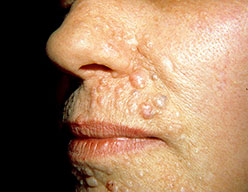
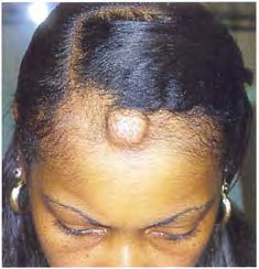
Tricholemmoma lesions usually present in adulthood. A tumor-suppressor gene (designated PTEN/MMAC1) has been demonstrated to play a causative role. Recently, some authors have proposed tricholemmoma to represent an old viral wart. Tricholemmoma (trichilemmoma) shows differentiation toward the outer root (trichilemmal) sheath of a normal hair follicle at the level of the bulb. Clinically, tricholemmomas are solitary, small (3 to 8 mm in diameter), asymptomatic, keratotic, or smooth-surfaced papules on the face, particularly around the nose and upper lip.Some cases arise in association with nevus sebaceus. Multiple facial tricholemmomas represent an important cutaneous marker for Cowden syndrome,. Patients with Cowden syndrome show a tendency to develop particular internal malignancies, especially involving the breast, colon, and thyroid.
Histologically
lesions show a lobular, folliculocentric, exo-endophytic proliferation of polygonal, pale-staining, glycogen-containing squamoid cells. The epidermal surface often reveals parakeratosis with scale crusts and sometimes a cutaneous horn (trichilemmal horn). The periphery of the lobules is bordered by columnar cells arranged in a palisade. Foci with small squamous eddies are occasionally observed within the center of tumor lobules. A conspicuous basement membrane usually surrounds the lesions. Desmoplastic tricholemmoma represents a special variant with prominently hyalinized or desmoplastic stroma.92 Treatment is by surgical excision.
|