| Raynaud’s phenomenon = ظاهرة رينود |
|
|
Raynaud Phenomenon
EPIDEMIOLOGY Studies of the epidemiology of Raynaud phenomenon are biased by under-reporting (most patients with primary Raynaud never seek medical attention) and studies done by investigators interested in the secondary forms of the disorder. Surveys indicate that Raynaud phenomenon affects up to 10 percent of the general population. Primary Raynaud phenomenon is estimated to be approximately twice as common as secondary Raynaud.3 Symptoms most often first develop in the teenage years, and most series show a female predominance of the disorder (female-male = 4:1). Increases in the frequency and severity of attacks during menses suggest that female sex hormones may be involved in the pathogenesis. Differences in seasonal skin temperatures, marital status, alcohol use, age, and smoking between women and men have also been suggested to contribute to the sex differences.5,6 Familial aggregation has been identified in a number of studies and suggests the contribution of genetic factors.7 Other associations reported in epidemiologic studies include living in a cold climate, occupation, cardiovascular disease, low body-mass index, and use of vibratory tools. ETIOLOGY AND PATHOGENESIS The pathophysiology of the vasospasm is complex and only partially understood. Studies show significant reductions of peripheral blood flow throughout all phases of central body cooling and re-warming, suggesting impairments of central thermoregulatory control mechanisms. The principal mechanisms thought to contribute to the development of Raynaud phenomenon include a local defect of a digital blood vessel, causing abnormal vascular reactivity or reduced blood flow; an enhanced localized production of vasoconstrictors or reduced production of vasodilators; hyperreactivity of the sympathetic nervous system; and abnormal properties of the blood that compromise distal perfusion. The results of gross histologic examinations of digital arteries in patients with primary Raynaud phenomenon are normal. Structural abnormalities of the digital microvasculature are frequently seen in patients with secondary forms of Raynaud phenomenon, particularly in the connective tissue diseases. Studies show a range of pathology, including intimal hyperplasia, narrowing or total occlusion of arteries, or thrombi. In most patients with systemic sclerosis, there exists evidence of activation and damage of the endothelium, fibrinolysis, and platelet activation. Microcirculatory flow studies with laser Doppler in patients with scleroderma have shown marked reductions in blood flow and hand temperature during an attack, with prominent abnormalities during re-warming—findings that suggest a failure of the arteriovenous anastomoses to open. Serotonin is also incriminated as an important mediator in the induction of ischemic attacks of Raynaud phenomenon. Patients have an increased sensitivity to intra-arterial infusions of serotonin, and S2-serotonergic antagonists relieve but do not prevent the induction of attacks. The evidence of direct central sympathetic nervous system hyperactivity is strongest in vibration-induced injury, in which the use of a vibration tool in one hand produces vasospasm in the other hand. Similarly, vasospasm can be inhibited by proximal nerve blockade in patients with vibration-induced injury. In general, most studies of the sympathetic nervous system in patients with primary or other secondary forms of Raynaud phenomenon have failed to detect evidence of sympathetic hyperactivity. The results of microelectrode studies of skin sympathetic nervous activity during cold pressor tests are normal, and plasma levels of catecholamines are not increased in the venous drainage of the hands of patients with Raynaud phenomenon.
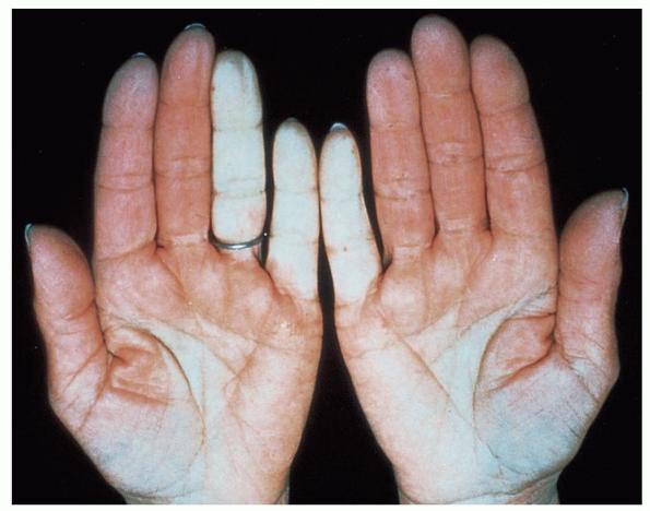
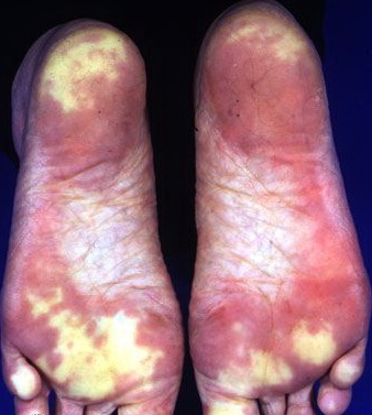
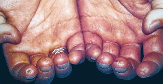
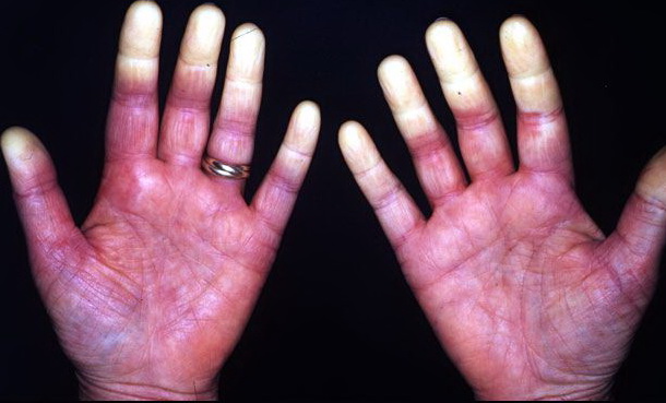
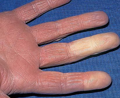
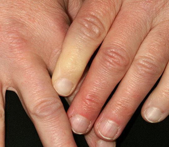
Consistent abnormalities in plasma fibrinogen, cold agglutinins, platelets, or cryoglobulins have not been demonstrated in primary Raynaud phenomenon, but are important in certain secondary cases, particularly scleroderma. Levels of von Willebrand's factor and soluble thrombomodulin, thromboxane B2 and β-thromboglobulin, and tissue plasminogen activator inhibitor-1 are increased in patients with scleroderma, when compared to levels in patients with primary Raynaud phenomenon. Genetic studies with microsatellite markers of extended families affected with primary Raynaud phenomenon have identified several potential candidate genes. CLINICAL FINDINGS A careful history and physical examination are important in the evaluation of a patient with suspected Raynaud phenomenon. History The history is important to elicit a clear description of the attacks and to screen for evidence of signs and symptoms suggestive of a secondary cause. Patients complain of episodic attacks of well-demarcated, white or blue digits induced by exposure to cold and sometimes by emotional stimuli . Often only a portion of the digit is affected, and the thumbs are typically spared. A classic tricolor change of pallor, cyanosis, and hyperemia described in most textbooks is rarely volunteered by patients; most describe only blanching of the digits accompanied by numbness. During the attacks, one or more fingers or toes may be numb and be described as “dead.” On re-warming, the digits may become bright red, and throbbing pain may occur. When pain is a prominent symptom in the ischemic phase, a secondary cause should be suspected. Attacks last minutes to hours. The fingers and toes are most commonly involved; however, the attacks may involve the nose, earlobes, or nipples. A careful review of systems is important to screen for symptoms of connective tissue disease (arthralgias, arthritis, dysphagia, heartburn, rash, photosensitivity, skin changes, muscle weakness, shortness of breath, or sicca), a drug-related etiology, symptoms of obstructive arterial diseases (intermittent claudication), and exposure to vibratory tools or continuous finger trauma. Cutaneous Lesions Attacks of Raynaud phenomenon, presumably stress induced, are commonly witnessed during the course of the history or physical examination. There is well-demarcated blanching or cyanosis of the digits extending from the tip to various levels of the digit . The digits distal to the line of ischemia are cold, while the proximal skin is pink and warmer. On re-warming, blanched digits may become cyanotic, because of the slow blood flow, and then bright red, because of reactive hyperemia. Persistent ischemic discoloration of digits suggests a secondary cause. The digits should be carefully examined for trophic or ischemic changes, which are signs of prolonged or severe attacks of Raynaud phenomenon. The skin may become atrophic, thin, and tight (sclerodactyly), with loss of hair over the dorsal surfaces. The nails may become brittle and deformed. Ulcerations, which can be extremely painful, particularly at night, may develop on the finger pads or around the nail bed. The ulcers heal slowly, leave characteristic small, pitted scars , and may become infected. Gangrene of the distal aspects of the digit is rare. Related Physical Findings The physical examination should pay attention to all pulses, and blood pressure should be obtained in both arms. Allen's test is useful to assess arterial and capillary function of the hands; abnormalities imply structural disease of the microcirculation and raise the suspicion of a secondary form of Raynaud phenomenon. The radial and ulnar arteries are simultaneously compressed by the examiner's thumbs while the patient opens and closes the fist to induce blanching of the palm. Selective arterial filling is judged by the rate of color return as pressure is sequentially released from the radial and ulnar arteries. Vascular
LABORATORY TESTS In all patients, a complete blood count, erythrocyte sedimentation rate, urinalysis, and anti-nuclear antibody test should be obtained. Additional laboratory studies should be directed by findings elicited by the history and physical examination. In patients with abnormal anti-nuclear antibody levels, tests for antibodies to specific nuclear antigens, such as Sm antigen or topoisomerase or centromere antigens, are helpful to detect early systemic lupus erythematosus or scleroderma . A routine chest radiograph should be obtained to look for a cervical rib or evidence of interstitial lung disease. Nail fold capillary microscopy is considered to be a useful procedure to distinguish primary from secondary Raynaud phenomenon. Although mild capillary abnormalities may be observed in patients with primary Raynaud phenomenon, patients with connective tissue diseases may have enlarged, deformed capillary loops surrounded by avascular areas . Serial studies reveal progressive decreases in the total number of nail fold capillary loops in secondary, but not primary, forms of Raynaud phenomenon. The patency of small arteries can be assessed by Doppler techniques. Digital subtraction arteriography should be reserved for selected patients with prolonged, severe ischemia for whom arterial reconstruction is a consideration. ▪ DIFFERENTIAL DIAGNOSIS Raynaud phenomenon is subdivided into primary (idiopathic) and secondary forms based on whether an underlying cause or disease association can be identified. Primary Raynaud Phenomenon Primary Raynaud phenomenon is a disorder in which known causes of attacks of peripheral vasospasm cannot be found. Criteria for the diagnosis of primary Raynaud phenomenon have been developed by Allen and Brown and LeRoy and Medsger. Several studies have examined the long-term outcome of patients with primary Raynaud phenomenon. Progression to a secondary form of Raynaud phenomenon, most commonly a connective tissue disease such as scleroderma, occurs in approximately 15 percent of patients during the first decade after onset. Variables predictive of a transition to a secondary form include nail fold capillary abnormalities, hand swelling, positive Allen's test, and anti-nuclear antibodies. Secondary Raynaud Phenomenon CONNECTIVE TISSUE DISEASES The connective tissue diseases are the most common cause of secondary Raynaud phenomenon . Among patients with scleroderma, 80 percent to 90 percent manifest Raynaud phenomenon and/or persistent vasospasm. It is the presenting symptom in about one-third of patients and may be the only manifestation of the disease for years. Raynaud phenomenon occurs in about one-third of patients with systemic lupus erythematosus, idiopathic inflammatory myopathies, and systemic vasculitis. Although patients with rheumatoid arthritis often complain of cold hands with mottled red and white areas, true Raynaud phenomenon appears to be no more common in persons with rheumatoid arthritis than in the general population. Arteriograms of patients with connective tissue diseases usually show digital and sometimes, ulnar or radial artery obstructions.
OCCUPATIONAL Raynaud phenomenon may be occupational in origin. It is especially common in individuals who use vibratory tools (e.g., air hammers, chain saws, rivet guns) and in those whose occupation requires prolonged exposure of the extremities to cold temperatures (e.g., butchers, ice cream workers, fish packers). Prevalence rates correlate with the vibration level of the tool and the duration of exposure; they can be as high as 90 percent in high-risk occupations such as logging or mining. NEUROLOGIC DISORDERS Any neurologic condition that produces permanent disuse of a limb can be associated with sympathetic nervous system disturbances to that limb. Patients often develop persistent vasospasm with coldness, paleness or cyanosis, and even ulcerations of the limb, and Raynaud phenomenon may occur. Thermoregulatory abnormalities may be
DRUGS AND TOXINS Propranolol, one of the most widely used β-adrenergic blockers for cardiovascular diseases and migraine headaches, is probably the most frequently used drug responsible for Raynaud phenomenon. Ergot preparations and methysergide used to treat migraine headaches may produce vasospasm. Intra-arterial use of many medications and recreational drugs can result in toxicity to endothelial cells with irreversible structural damage to the microvasculature of the extremities and be responsible for severe Raynaud phenomenon. The chemotherapeutic agents bleomycin and vinblastine also may cause the phenomenon.22 HYPERVISCOSITY Patients with hyperviscosity from cryoglobulinemia, macroglobulins, cold agglutinins, and polycythemia can exhibit Raynaud phenomenon. Cryoglobulins are most commonly present in patients with multiple myeloma, but they also occur in patients with rheumatic diseases, chronic infections, leukemia, and lymphoma, and as an idiopathic condition. MISCELLANEOUS The most common endocrine disturbance associated with Raynaud phenomenon is hypothyroidism; symptoms usually remit with thyroid hormone replacement. Raynaud phenomenon may be a feature of various infectious disorders, including subacute bacterial endocarditis, Lyme disease, and viral hepatitis, presumably a reflection of systemic vasculitis. Peripheral vasospasm may also occur in association with malignant tumors, including pheochromocytoma, carcinoid, and ovarian carcinoma. The major forms of vascular ischemia that must be distinguished from Raynaud phenomenon are cold digits, chilblain (pernio), livedo reticularis, and acrocyanosis (see Chap. 93). Many patients complain of cold, sometimes painful digits without color changes. This condition likely represents one extreme of the spectrum of normal sympathetic nervous system activity. Chilblain is an inflammatory condition of the skin of the extremities induced by cold. Patients develop a bluish-red discoloration and edema, typically involving the lower limb and associated with warmth, erythema, and burning. In severe cases, hemorrhagic lesions, bullae, or ulcers may develop, and secondary infections may supervene. The lesions last from 7 to 10 days, often leaving a residual pigmentation of the skin. Livedo reticularis is a bluish discoloration of the skin of the extremities with a characteristic lacy, irregular appearance. The bluish discoloration becomes more intense on exposure to cold and may disappear in a warm environment. Most patients are entirely asymptomatic, although livedo reticularis may be a feature of the anti-phospholipid syndrome, in which patients are at increased risk for venous and arterial thromboses, thrombocytopenia, and pregnancy losses. In acrocyanosis, the hands, and, less commonly, the feet, develop a persistent bluish discoloration. The blue color is intensified by exposure to cold and is converted into a purplish or red color by warming; a pallor phase is absent. The skin is cold, and the palms are often wet and clammy from sweat. Trophic changes or ulcerations are rarely observed. TREATMENT The management of Raynaud phenomenon is guided by the frequency and severity of attacks and the complications from ischemia . Secondary forms of Raynaud phenomenon require treatment directed at the underlying medical disorder, discontinuation of drugs causing the vasospasm, or occupational modifications. General Measures Mild Raynaud phenomenon is generally easy to control with lifestyle changes to minimize exposure to the cold; dressing warmly with loose-fitting, layered clothing; and keeping the thermostat a few degrees higher than normal. Limiting time spent outdoors in winter, wearing insulated gloves, and using hand or foot warmers are usually helpful. Patients should be taught to recognize and terminate attacks promptly by returning to a warmer environment and applying local heat to the hands (e.g., by placing their hands in warm water or by using a hair dryer). Patients should be strongly encouraged to stop smoking and to avoid
Digital ulcers from Raynaud phenomenon can be extremely painful and typically take weeks or months to heal completely. Pain control is an important part of therapy, because pain can lead to additional vasospasm and more ischemia. On occasion, narcotic pain medications may be necessary to control symptoms. The finger should be soaked in a tepid antiseptic solution (e.g., half-strength hydrogen peroxide) twice daily to soften or loosen the crust or eschar. After drying, an antibiotic ointment is applied to the ulcer, and the digit is covered with an occlusive dressing. Maximum drug therapy with a calcium channel blocker should be used throughout treatment. Infection is a common complication of digital ulcers and is typically manifest by increasing pain, erythema, swelling, or purulent drainage. Cultures usually demonstrate Staphylococcus sp., and treatment with dicloxacillin or cephalosporins is usually effective. Drug Therapy Various drugs have been used to treat Raynaud phenomenon, including vasodilators, platelet inhibitors, serotonin antagonists, and fibrinolytics. Drug therapy is usually reserved for patients with prolonged or frequent attacks that fail to respond to conservative measures. In general, improvements with drug therapy are more pronounced in patients with primary Raynaud phenomenon, presumably as a consequence of fixed, structural damage in patients with secondary forms. Sympatholytic drugs, including reserpine, methyldopa, phenoxybenzamine, and tolazoline, have been used in management, although they have not been well studied. In a pilot study, the selective serotonin reuptake inhibitor fluoxetine reduced the frequency and severity of Raynaud attacks. Additional drug therapies reported to be beneficial in the treatment of Raynaud phenomenon include low molecular weight heparin, prazosin, the angiotensin II receptor antagonist losartan, and stanozolol.29 The calcium channel blockers are by far the most widely used and effective drugs for treating Raynaud phenomenon. Vasodilating properties vary among different agents. Nifedipine (10 to 20 mg tid or qid) reduces the severity and frequency of attacks; the long-acting preparation of nifedipine is better tolerated but may be less effective. Diltiazem (60 mg tid or qid) may be substituted if nifedipine is ineffective or not well tolerated. Side effects of calcium channel blockers include fluid retention, light-headedness, and heartburn; these may limit therapy. Topical nitroglycerin paste (2 percent) and a sustained-release transdermal glyceryl patch are helpful in selected patients. Intravenous prostaglandin E1 and prostacyclin (PGI2) and iloprost (a PGI2 analog) have been shown to have beneficial effects in patients with severe Raynaud phenomenon; however, the vasodilatory effects are not sustained and long-term therapy is required. Oral prostaglandins are currently available only as investigational agents. Sympathectomy Sympathectomy may be a consideration for the management of patients with refractory, disabling attacks or with an acutely ischemic digit that is unresponsive to other measures. A positive vasodilator response to a stellate ganglion block or epidural infusion should be documented before a permanent procedure is done. Lumbar sympathectomy has an important role in the management of severe Raynaud phenomenon of the feet, and selective digital sympathectomy may be used to relieve pain and heal digital ulcers in patients with ischemic digits. The long-term effects of sympathectomy are poor, with up to two-thirds of patients reporting no benefit at the end of 1 year. Newer procedures for thoracic sympathectomy, including percutaneous radio frequency36 and thoracoscopic procedures,37 have been advocated as technically easier to perform with fewer complications and more promising long-term results. Severe Vasospasm Severe vasospasm with prolonged ischemia (dead-white finger) poses a threat of gangrene and amputation; it is considered a medical emergency. The patient should be hospitalized and the affected extremity put to rest. Nifedipine (10 to 20 mg tid) should be started immediately, as well as prostaglandin E1 (6 to 10 ng/kg/minute) or PGI2 (0.5 to 2 ng/kg), given by continuous intravenous infusion for several hours over 3 consecutive days. Intra-arterial phentolamine or tolazoline may reverse acute vasospasm, but monitoring of vital signs is essential, and these drugs need to be used with great caution. A digital (or stellate ganglion) block with lidocaine hydrochloride or bupivacaine hydrochloride (without epinephrine) relieves pain and produces a chemical sympathectomy that may reverse vasoconstriction. Sympathectomy (thoracic, lumbar, or digital) should be considered in patients who have a positive response. Arterial reconstruction should be reserved for patients with angiographically documented occlusive vascular disease.
|