|
Pemphigus erythematosus (Senear Usher syndrome)= الفقاع الحمامي --سينيرأوشر |
 |
 |
 |
PEMPHIGUS
ERYTHEMATOSUS
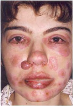
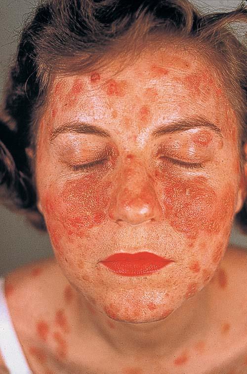
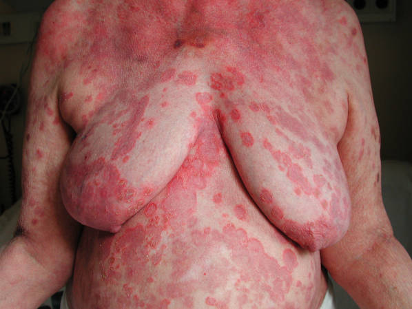
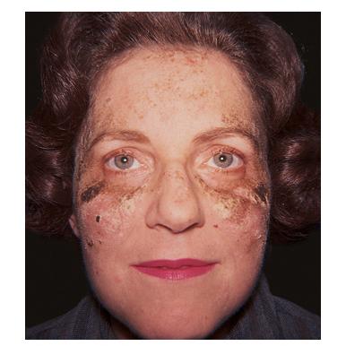
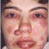
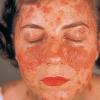
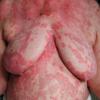
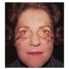
Also known as Senear-Usher syndrome, pemphigus erythematosus is simply the localized form of PF. Typical scaly and crusted lesions of PF occur across the malar area of the face and in other seborrheic areas. Pemphigus erythematosus may remain localized for years, or it may evolve into more generalized PF. If there is a unique aspect of pemphigus erythematosus, it is the immunofluorescence findings noted in Immunopathology.In addition, many patients with pemphigus erythematosus show serologic findings suggestive of systemic lupus erythematosus, especially the presence of anti-nuclear antibodies, although few patients have been reported to actually have the two diseases concurrently.
|
Histopathology.
The light microscopic features are identical to those of pemphigus foliaceus (Fig. 9-12). Interface dermatitis may also be apparent in rare cases, making distinction from lupus erythematosus difficult.
|
|
IF Testing. DIF testing of perilesional skin reveals squamous intercellular substance deposition of IgG in >75% of cases and granular deposition of IgM and IgG (l.e., a positive lupus band test) at the dermal-epidermal junction. IIF study using monkey esophagus as substrate reveals squamous intercellular substance deposition of IgG in 80% of cases. Antinuclear antibodies are observed in 30% to 80% of cases.
|
|
Ultrastructural Study. Pemphigus erythematosus is identical to pemphigus foliaceus in its ultrastructural alterations.
|
|
Differential Diagnosis. The differential diagnosis is the same as in pemphigus foliaceus. The presence of interface dermatitis in some cases leads to confusion with lupus erythematosus and PNP. Subcorneal acantholysis is not a feature of lupus erythematosus.
|
|