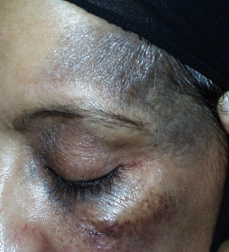


Nevus of Ota
Nevus of Ota (nevus fusco-caeruleus ophthalmomaxillaris) was first described by Ota
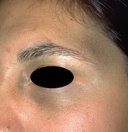
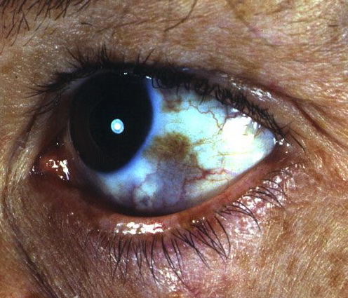
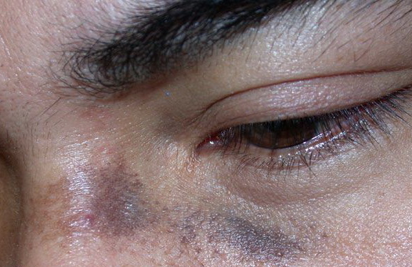
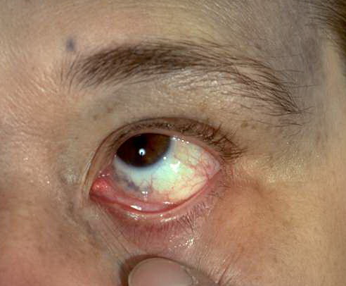
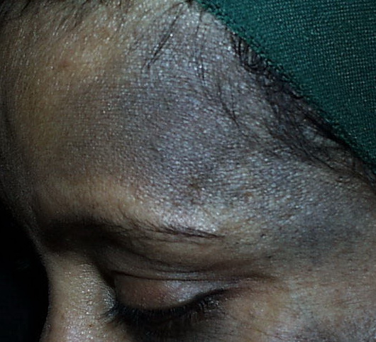
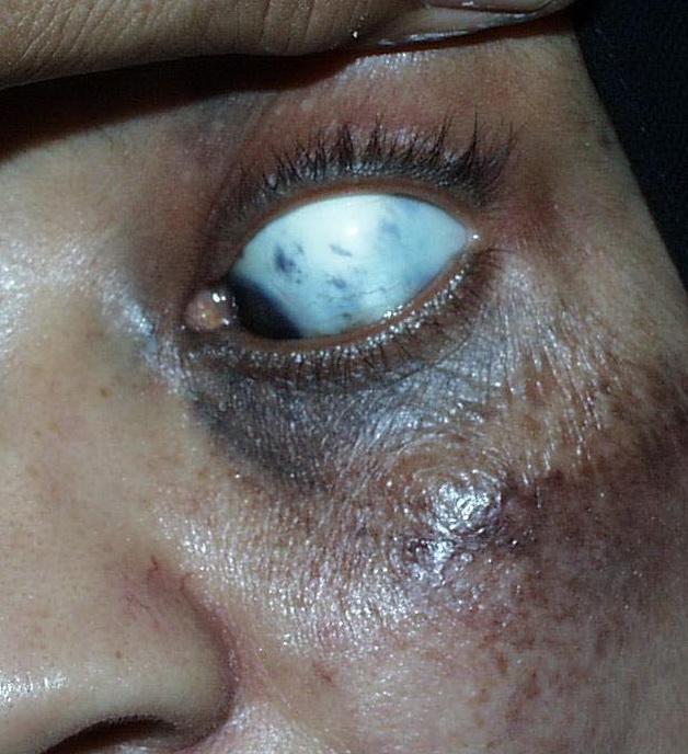
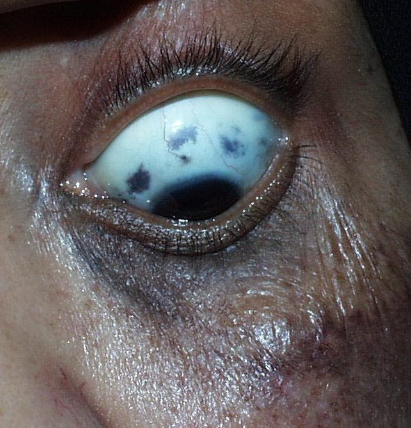
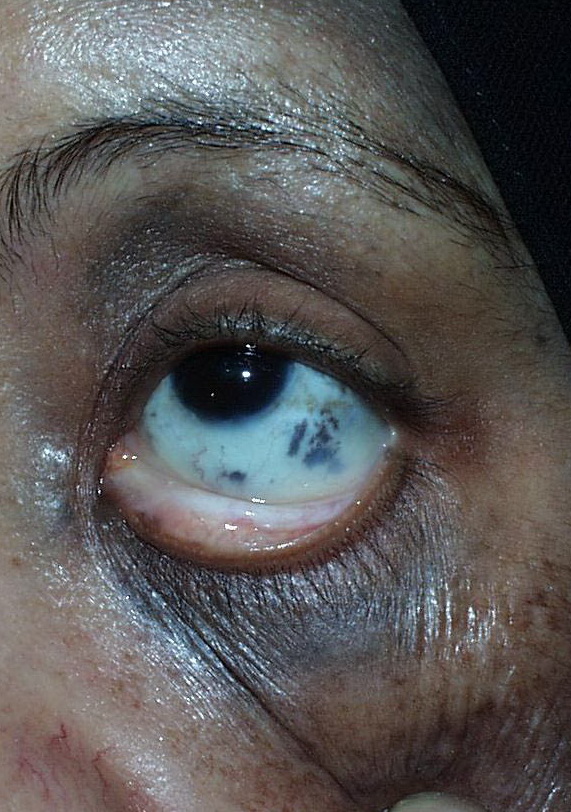
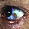
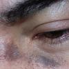
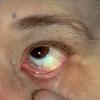
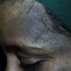
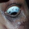
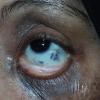
in 1939. It is characterized by blue-black or gray-brown dermal melanocytic pigmentation and typically occurs in areas innervated by the first and second branches of the trigeminal nerve. Mucosal pigmentation may occur, involving the conjunctiva, sclera, and tympanic membrane (oculodermal melanocytosis) . It is most frequently seen in the Asian population, has a female predominance, and is usually congenital, although appearance in early childhood or in puberty has been described. In 1988, Ota nevus was subclassified as mild (type 1), moderate (type 2), intensive (type 3), and bilateral (type 4). Bilateral Ota nevus should be differentiated from Hori nevus (acquired bilateral nevus of Ota-like macules), which is acquired, does not have mucosal involvement, and is less pigmented .
Malignant melanoma may rarely develop in a nevus of Ota. This necessitates careful follow-up of the lesion, especially if it occurs in Caucasian patients, in whom malignant degeneration seems to be more frequent. Malignant melanocytic tumors in association with nevus of Ota have been shown to arise in the chorioidea, brain, orbit, iris, ciliary body, or optic nerve.