
Acne Rosacea
Rosacea is a common condition characterized by symptoms of facial flushing and a spectrum of clinical signs, including erythema, telangiectasia, coarseness of skin, and an inflammatory papulopustular eruption resembling acne.
Definition and subtypes
An expert committee assembled by the National Rosacea Society explicitly defined and classified rosacea in April 2002 into 4 different subtypes based on specific clinical signs and symptoms. This categorization was an important step in the treatment of rosacea. Currently, the therapeutics of rosacea empirically target the signs and symptoms of the disease because investigators do not understand the details of its pathophysiology. Therefore, this classification system aides clinicians in treatment by highlighting the preponderance of one or more of the clustering signs of presentation and, thus, helps to specify which therapeutic approach to initiate.
The diagnosis of rosacea is a clinical diagnosis. Skin biopsy may be necessary to exclude other disease states that mimic the clinical presentation of rosacea. For example, the clinician must exclude polycythemia vera, connective-tissue diseases (eg, lupus erythematous, dermatomyositis, mixed connective-tissue disease), photosensitivity, carcinoid syndrome, mastocytosis, long-term application of topical steroids, contact dermatitis, and photosensitivity before making the diagnosis of rosacea.
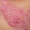
Rosacea is defined by persistent erythema of the central portion of the face lasting for at least 3 months. Supporting criteria include flushing, papules, pustules, and telangiectasias on the convex surfaces. Secondary characteristics are burning and stinging, edema, plaques, a dry appearance, ocular manifestations, and phymatous changes. The prevalence of these findings designates the subclassification of the presentation and, additionally, the therapeutic options.1,2,3
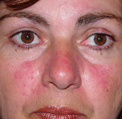
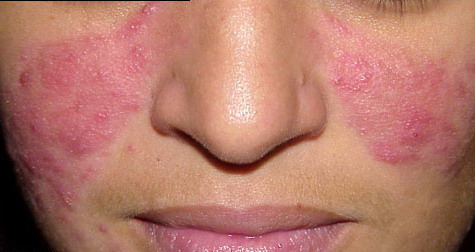
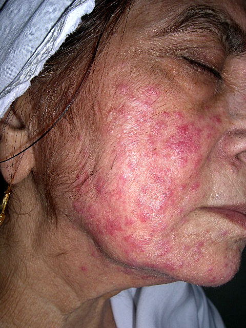
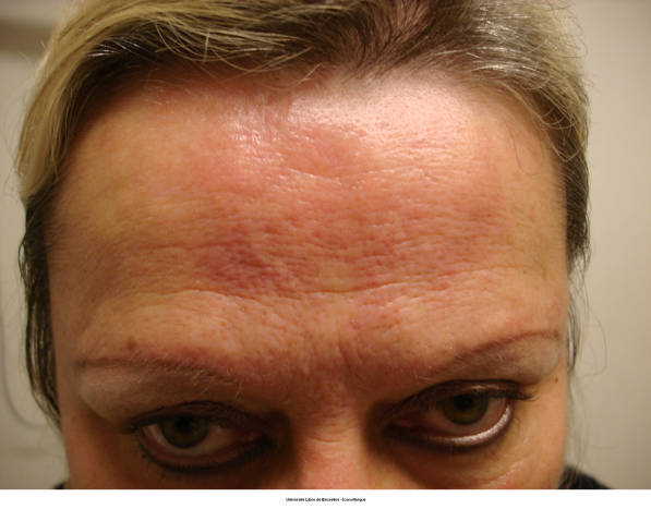
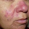
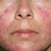
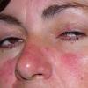
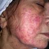
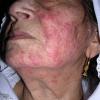
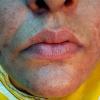
Erythematotelangiectatic type
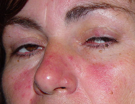
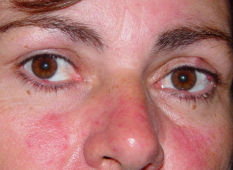
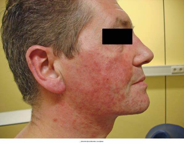
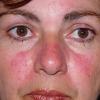
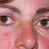
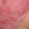
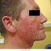
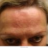
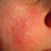
Central facial flushing, often accompanied by burning or stinging, is the predominant sign in erythematotelangiectatic rosacea (ETR). The redness usually spares the periocular skin. These patients typically have skin with a fine texture that lacks a sebaceous quality characteristic of other subtypes. The erythematous areas of the face at times appear rough with scale likely due to chronic, low-grade dermatitis. Frequent triggers to flushing include acutely felt emotional stress, hot drinks, alcohol, spicy foods, exercise, cold or hot weather, and hot baths and showers. These patients also report that the burning or stinging is exacerbated when topical agents are applied.
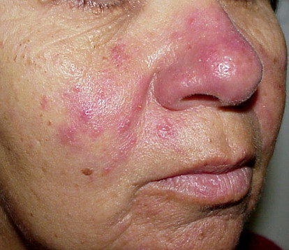
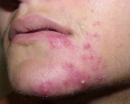
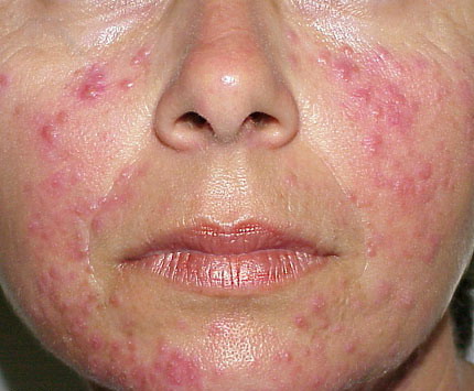
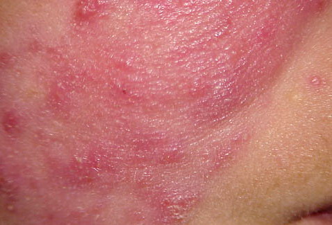
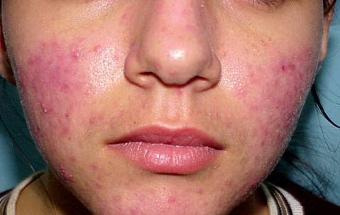
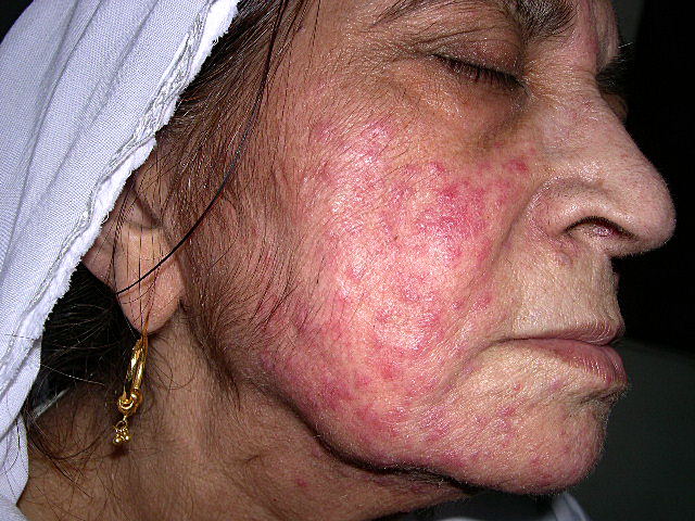
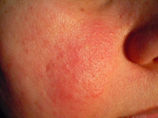
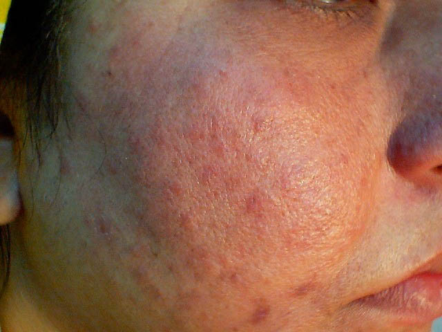
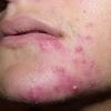
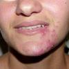
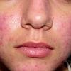
Papulopustular rosacea
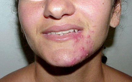
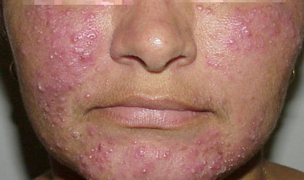
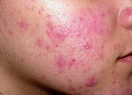
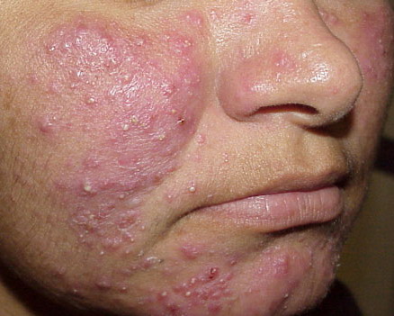
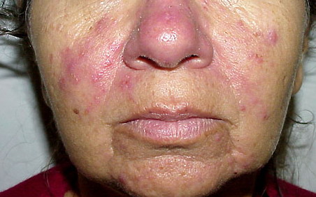
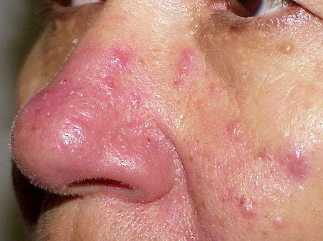
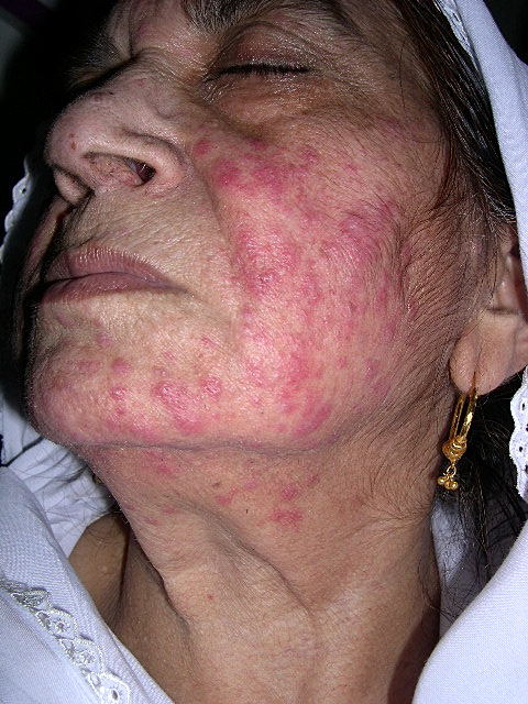
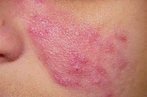
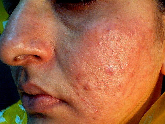
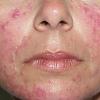
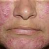
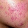
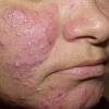
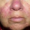
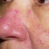
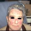
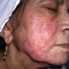
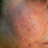
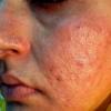
Papulopustular rosacea (PPR) is the classic presentation of rosacea. Patients are women of middle age who predominately present with a red central portion of their face that contains small erythematous papules surmounted by pinpoint pustules. One may elicit a history of flushing. Telangiectasias are likely present but may be difficult to distinguish from the erythematous background in which they
exist
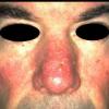
Phymatous rosacea
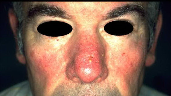
Phymatous rosacea is defined as marked skin thickenings and irregular surface nodularities of the nose, chin, forehead, one or both ears, and/or the eyelids. Four distinct histologic variants can occur with rhinophyma (associated changes of the nose) that include glandular, fibrous, fibroangiomatous, and actinic. The mainstays of treatment are isotretinoin topical application and surgical correction. This varies from other rosacea subtypes.
Ocular rosacea
Ocular manifestations may precede the cutaneous signs by years. Yet, frequently they develop concurrently with dermatologic manifestations. The ocular manifestations include blepharitis, conjunctivitis, inflammation of the lids and meibomian glands, interpalpebral conjunctival hyperemia, and conjunctival telangiectasias. Patients may describe eye stinging or burning, dryness, irritation with light, or foreign body sensation. Ocular rosacea, similar to phymatous rosacea, has a distinct therapeutic management. Therefore, dermatologists must ask their patients specifically about ocular symptoms and perform a thorough physical examination to rule out this type of rosacea.
Pathophysiology
The etiology of rosacea is unknown. However, several factors, such as vasculature, climatic exposures, dermal matrix degeneration, chemicals and ingested agents, pilosebaceous unit abnormalities, microbial organisms, ferritin expression, reactive oxygen species (ROS), increased neoangiogenesis, and dysfunction of antimicrobial peptides (AMPs), likely play a role in its development.4 Furthermore, the distinct subtype of rosacea is likely determined by a patient's unique sensitivity to these triggers.
Vasculature
Increased blood flow to the blood vessels of the face and increased numbers of blood vessels that are closer to the surface of the face are thought to be responsible for the redness and flushing associated with rosacea. Furthermore, vasodilatation, the normal response to hyperthermia, is thought to be more pronounced or exaggerated in those individuals with rosacea.
Climatic exposures
Some evidence suggests that harsh climatic exposures damage cutaneous blood vessels and dermal connective tissue. This also includes exposure to solar irradiation, which may explain why rosacea predominately affects the facial convexities and has a tendency to flare in the spring. However, other studies suggest the contrary, in that most patients' symptoms do not worsen in the sunlight and do not flare with an acute exposure to ultraviolet (UV) light.
Dermal matrix degeneration
Rosacea involves associated damage to the endothelium and degeneration of the dermal matrix. However, it is not known whether the initial damage is in the dermal matrix and this leads to poor tissue support of cutaneous vessels, causing pooling of serum, inflammatory mediators, and metabolic waste, or whether the initial abnormality exists in the cutaneous vasculature and this leads to leaky vessels and delayed clearance of serum proteins, inflammatory mediators, and metabolic waste, thus resulting in matrix degeneration.
Chemicals and ingested agents
Spicy foods, alcohol, and hot beverages may trigger a flushed face in patients with rosacea. However, most evidence does not support dietary factors playing a central role in the pathogenesis. Moreover, certain medications, such as amiodarone, topical steroids, nasal steroids, and high doses of vitamins B-6 and B-12, may cause flares for patients with rosacea.
Perivascular versus perifollicular inflammation
An inflammatory infiltrate may exist in a perivascular and/or a perifollicular location; however, evidence is conflicting regarding which location predominates. To answer this question, more studies need to be designed to categorize subtypes of rosacea because the answer varies depending on the subclassification.
Microbial organisms
Demodex species (mites that normally inhabit human hair follicles) may play a role in the pathogenesis of rosacea. Some studies suggest that Demodex prefers the skin regions that are affected in rosacea, such as the nose and cheeks.5 Research also supports that an immune response of helper-inducer T-cell infiltrates occurs, surrounding the Demodex antigens in patients with rosacea. Yet, conflicting evidence indicates that Demodex does not induce an inflammatory response in patients with rosacea. Moreover, Demodex is found in large numbers of healthy individuals without rosacea. More studies need to be performed to determine whether Demodex truly is pathogenic.
Additionally, inconclusive evidence suggests that Helicobacter pylori is associated with the etiology of rosacea. However, many of the studies have not controlled for confounding variables that influence H pylori prevalence, such as sex, age, socioeconomic status, and medications. Furthermore, these studies were not statistically powered to account for the ubiquitous nature of H pylori infection.
Ferritin expression
Iron catalyzes the conversion of hydrogen peroxide to free radicals, which leads to tissue injury by damaging cellular membranes, proteins, and DNA. At the cellular level, iron that is not metabolized is stored as ferritin. In a 2009 study, skin biopsy specimens from patients with rosacea were immunohistochemically analyzed, and the number of ferritin-positive cells was significantly higher in affected individuals compared with control subjects. Additionally, higher ferritin positivity correlated with more advanced subtypes of rosacea. Thus, increased release of free iron from proteolysis of ferritin can result in oxidative damage to the skin, which may contribute to the pathogenesis of rosacea.6
Reactive oxygen species
Early in the inflammatory process, ROS are released by neutrophils, which are postulated to have a central role in the inflammation associated with rosacea. Free radicals, such as superoxide anions and hydroxyl radials, in addition to other reactive molecules, such as molecular oxygen, singlet oxygen, and hydrogen peroxide, comprise many of the ROS that lead to oxidative tissue damage. Several mechanisms explain how ROS result in skin inflammation, most notably the deactivation of natural defenses caused by excessive oxidant stress from ROS; chemical and oxidative modification of proteins and lipids by ROS; alteration of the lipid balance in rosacea patients, which, in normal proportions would suppress the creation of ROS; production of cytokines and other inflammatory mediators by keratinocytes, fibroblasts, and endothelial cells damaged by ROS; and the generation of ROS by cathelicidins, which are found in greater amounts in the facial skin of affected individuals.7
Neoangiogenesis and vascular endothelial growth factor (VEGF) overexpression
Studies performed using video capillaroscopy on erythematotelangiectatic rosacea lesions showed increased neoangiogenesis and blood vessel enlargement. Multiple immunohistochemistry studies showed increased VEGF expression in vascular endothelium in lesional versus nonlesional skin of rosacea patients. Cuevas et al8 used topical dobesilate, an inhibitor of angiogenic growth factor, for the treatment of erythematotelagiectatic rosacea and reported an improvement in erythema and telangiectasia after 2 weeks.4
Antimicrobial peptides
AMPs are small molecular weight proteins that are a part of the innate immune response and have demonstrated broad-spectrum antimicrobial activity against bacteria, viruses, and fungi. They are rapidly released upon injury and/or infection of the skin, and they have been implicated in the pathogenesis of many inflammatory skin diseases. Cathelicidins and β-defensins are 2 well-known types of AMPs, of which the former has been shown to be expressed in abnormally high levels in patients with rosacea.
Specifically, the LL-37 peptide form of cathelicidin, in addition to proteolytically processed forms of LL-37, have been found in significantly different amounts in rosacea patients compared with healthy individuals. LL-37 is expressed by polymorphonuclear leukocytes and lymphocytes. LL-37 interacts with endothelial cells and stimulates angiogenesis both in vitro and in vivo. It also modulates the expression of VEGF.4 Injection of LL-37 and these novel peptides derived from LL-37 into mice induced inflammation, erythema, and telangiectasia; therefore, researchers hypothesized that an excess of cathelicidins coupled with abnormal processing caused diseas
History
Patients are likely to have a background of facial flushing, often dating to childhood or the early teens. In adult life, flushing may be increasingly precipitated by hot drinks, heat, emotion, and other causes of rapid body temperature changes. Some patients report flushing with alcohol, which is not specific.
The symptoms are usually intermittent but can progressively lead to permanently flushed skin. The latter may be described as high color and is associated with the development of permanent telangiectasia. Additionally, a few individuals report a gritty quality of the eyes and facial edema.
Physical
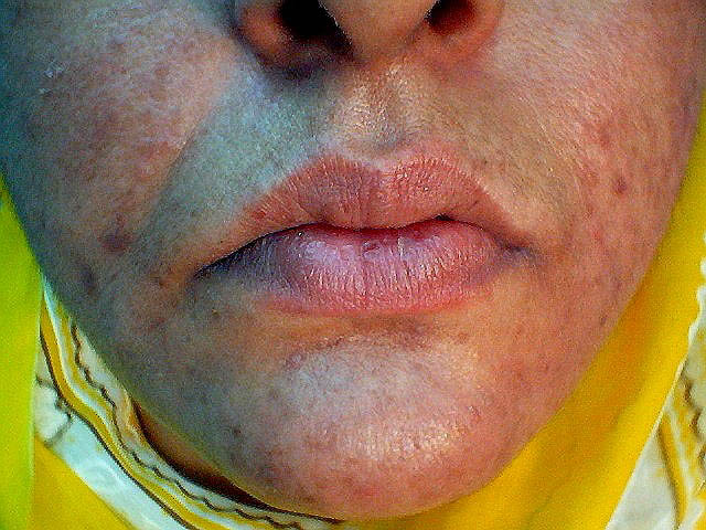
The disease consists of a spectrum of symptoms and signs, with most patients failing to develop every stage of disease. Variable erythema and telangiectasia are seen over the cheeks and the forehead. Inflammatory papules and pustules may be predominantly observed over the nose, the forehead, and the cheeks. Extrafacial involvement uncommonly occurs over the neck and the upper part of the chest. Prominence of sebaceous glands may be noted, with the development of thickened and disfigured noses (rhinophyma) in extreme cases. Unlike acne, patients generally do not report greasiness of the skin; instead, they may experience drying and peeling. The absence of comedones is another helpful distinguishing feature. Ocular lymphedema may be prominent but is uncommon. The condition generally does not produce scarring.
Rhinophyma may occur as an isolated entity, without other symptoms or signs of rosacea. Rhinophyma can be disfiguring and therefore distressing for patients. Some authorities consider rhinophyma to represent a different disease process.10
Lymphedema may be marked periorbitally, and, on occasion, it is the presenting symptom.
Symptoms of ocular rosacea may be accompanied by conjunctival injection, and, rarely, chalazion and episcleritis may occur.
Rosacea fulminans (pyoderma faciale) is fortunately a rare complication and is characterized by the development of nodules and abscesses with sinus tract formation accompanied by systemic signs.
Both seborrhea and seborrheic dermatitis/blepharitis are not uncommonly observed in patients with rosacea. The reasons for these associations are not well understood.
A rare caseating granulomatous variant of rosacea (acne agminata/lupus miliaris disseminatus faciei) can manifest with inflammatory erythematous or flesh-colored papules distributed symmetrically across the upper part of the face, particularly around the eyes and the nose. The lesions tend to be discrete, and surrounding erythema is not a marked feature but may be present. This pattern of rosacea is sometimes associated with scarring and may be resistant to conventional treatment
Causes
A rosacealike syndrome (including perioral dermatitis) can result from the indiscriminate use of potent corticosteroids on the face. A number of aggravating factors may be recognized. Excess wind and UV light (weathering) exposure may accelerate the disease process
Other Problems to Be Considered
The differential diagnosis largely depends on the pattern of rosacea. Erythematotelangiectatic rosacea (ETR) can resemble seborrheic dermatitis, lupus erythematosus, and other photodermatoses. Carcinoid syndrome and mitral valve incompetence are overlooked causes of erythema and telangiectasia. Acneiform rosacea may be simulated by acne, bromoderma and iododerma, perioral dermatitis, and pustular folliculitis. Acne agminata can be indistinguishable from lupus vulgaris and cutaneous sarcoidosis
Procedures
A skin biopsy is sometimes performed to exclude other cutaneous diseases, such as lupus or sarcoidosis.
Histologic Findings
The histologic features of rosacea depend on the stage of disease. Nonpustular lesions show a nonspecific perivascular and perifollicular lymphohistiocytic infiltrate, accompanied by occasional multinucleated cells, plasma cells, neutrophils, and eosinophils. Papulopustular lesions demonstrate more pronounced granulomatous inflammation and sometimes perifollicular abscesses. Demodex folliculorum may be abundant in nearby follicles. The histologic features of acne agminata are striking, demonstrating caseating granulomata with negative stains for mycobacteria and fungi. See the images below.
.
Medical Care
Before the initiation of therapy, the triggering factors that exacerbate the patient's rosacea should be identified and avoided if possible. These factors may be unique to each individual patient. Common triggering factors include hot or cold temperatures, wind, hot drinks, caffeine, exercise, spicy food, alcohol, emotions, topical products that irritate the skin and decrease the barrier, or medications that cause flushing.11,12 Some patients find that regular facial massage reduces lymphedema. Rosacea fulminans is treated with moderately high doses of prednisolone (30-60 mg/d) followed by oral isotretinoin.
Sunscreen13
The use of daily broad-spectrum sunscreen is recommended for all patients with rosacea. A sunscreen that protects against both UV-A and UV-B light should be selected. Physical blockers such as titanium dioxide and zinc oxide are well tolerated. Additionally, the sunscreen should contain protective silicones such as dimethicone or cyclomethicone. Green-tinted sunscreens can provide coverage of the erythema.
The patient is encouraged to avoid astringents, toners, menthols, camphor, waterproof cosmetics requiring solvents for removal, or products containing sodium lauryl sulfate.
Laser14
Nonablative laser is effective against rosacea by remodeling of the dermal connective tissue and improving the epidermal barrier. The major disadvantage of this therapy is its cost because it is not covered by insurance. It requires 1-3 treatments 4-8 weeks apart to achieve the best results.
Vascular lasers are the mainstay of rosacea therapy. These include pulsed dye laser (585 or 595 nm), the potassium-titanyl-phosphate laser (532 nm), and the diode-pumped frequency-doubled laser (532 nm). These wavelengths allow selective absorption by oxyhemoglobin, leading to vessel reduction with minimal damage to surrounding tissue or scarring. To be effective against deeper facial vessels, longer wavelengths of lasers are required, including the diode laser (810 nm), the long-pulsed Alexandrite laser (755 nm), and the long-pulsed Nd:YAG laser (1064 nm).
Intense pulsed-light therapy is a multichromatic laser with different targets, including melanin and hemoglobin. Therefore, it is also useful for facial rejuvenation, affecting vascular lesions, pigmented lesions, and hair.
Surgical Care
Permanent telangiectasia may be treated by electrosurgery or the 585-nm pulsed dye laser. However, facial erythema is not improved, and new telangiectasias develop with the passage of time. Cosmetic improvement of rhinophyma may be produced by mechanical dermabrasion, carbon dioxide laser peel, and surgical shave techniques.
Diet
Dietary modulation should aim at avoidance of triggers.