Angioma serpiginosum
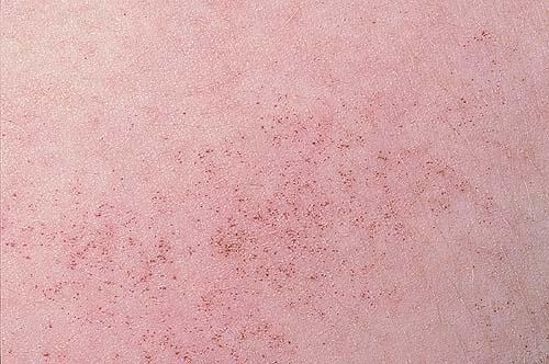
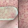
Angioma serpiginosum is an uncommon cutaneous vascular nevus of superficial capillaries characterized by minute puncta in clusters or in a linear array (a serpiginous pattern). These puncta result from a congenital hyperplasia or ectasia of preexisting superficial dermal capillaries, which may ultimately disappear (probably as a result of thrombosis). Electron microscopic findings have supported the view that these lesions are due to a vascular anomaly rather than a simple telangiectasia.
Clinical
History
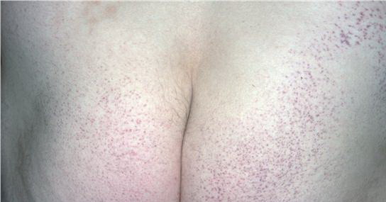
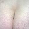
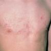
Angioma serpiginosum, a rare vascular nevoid disorder due to ectatic dilation of capillaries in the papillary dermis, is found almost exclusively in females. In 2005, Sandhu and Gupta7 reported 2 rare cases—one with familial involvement and the other with an extensive distribution of lesions. Affected individuals tend to have grouped erythematous punctate lesions on the lower limbs or buttocks. Note the following:
- A port-wine stain may be the first evidence of this disorder, appearing during the first few months of life.
- Years later, it may slowly enlarge, not by a uniform edge but rather by minute satellites ranging from copper-red to vividly red.
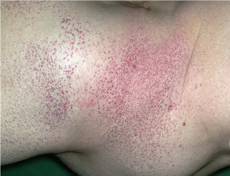
- Satellites spread into circles and gradually coalesce, producing the irregular serpiginous pattern.
- When the lesion resolves, it does so with a very superficial scar.
- Lesions can be located anywhere on the body and have been reported in all areas except the palms and mucous membranes.7
- Areas of predilection are the extremities, especially the lower extremities.
- Patches are progressive and asymptomatic and rarely resolve.
- Rarely, patches may be extensive in distribution.7
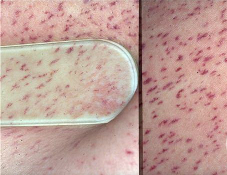
- Numerous small, relatively well-demarcated, round-to-oval red lagoons may be visualized with dermoscopy, which can be beneficial in the diagnosis of angioma serpiginosum.
- Retinal involvement has been described.
- Angioma serpiginosum with esophageal papillomatosis has been described as an X-linked dominant condition that maps to Xp11.3-Xq12.10 A 4-generation family with localized subepidermal telangiectasias following Blaschko lines (angioma serpiginosum) was described, with vascular streaks present at birth and that progressed slowly thereafter. Several family members had papillomatosis of the entire esophagus.
Physical
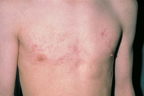
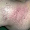
Angioma serpiginosum is composed of reddish-purple puncta that may be as large as 1 mm. They are usually found grouped on the lower extremities in a serpiginous pattern. Rarely, the sole may be involved. Punctate erythematous maculae on the backs of the hands, arms, and shoulders may appear following a pregnancy.
Angioma serpiginosum is variably compressible. The lack of inflammation, hemorrhage, or hemosiderin pigmentation is characteristic. Diascopic pressure applied to the lesion may produce only partial emptying, with some small tufts distended by purple venous blood remaining unchanged.
Histologic Findings
The overlying epidermis is normal. The dermal papilla and subpapillary regions of the dermis show dilated capillaries with a thickening of the capillary walls. No inflammatory changes, hemorrhage, or hemosiderin depositions are present. Ultrastructural analysis of several lesions has shown that some thickening of vessel walls may occur from a heavy precipitate of basement membrane–like, fine fibrillar material mixed with thin collagen fibers. Some of these dilated capillaries show slitlike protrusions of lamina into the endothelial lining.
Treatment
Medical Care
This vascular lesion does not require medical care.
Surgical Care
Electrolysis or laser surgery of an individual lesion may be beneficial. Good cosmetic results can be achieved with a tunable pulse dye laser by selective photothermolysis of the vascular ectasias.12,18,19 With the tunable pulse dye laser, good-to-excellent results may be achieved in 4 or fewer visits